
Fig. 3
- ID
- ZDB-FIG-151008-17
- Publication
- Hu et al., 2015 - Loss of DDB1 Leads to Transcriptional p53 Pathway Activation in Proliferating Cells, Cell Cycle Deregulation, and Apoptosis in Zebrafish Embryos
- Other Figures
- All Figure Page
- Back to All Figure Page
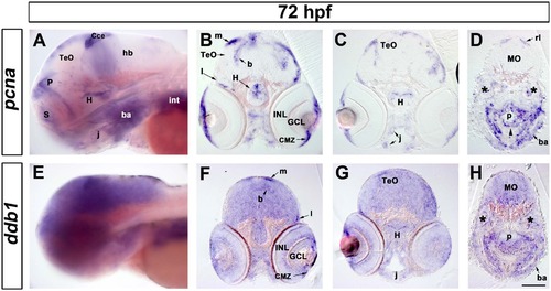
Expression of ddb1 in proliferation regions in wild type larvae at 72 hpf. (A-H) Characterization of pcna (A-D) and ddb1 (E-H) expression in wild type larvae at 72 hpf. Lateral views (A, E) and sections (20 µm; B-D, F-H). (A, E) ddb1 was broadly expressed in the brain while pcna was restricted to proliferation zones. (B-D, F-H) In the medial (m), lateral (l) and basal (b) proliferation zones of the tectum opticum (TeO) and the hypothalamic proliferation zone (H), elevated expression of ddb1 was detected compared to other areas of the brain. ddb1 was also highly expressed in the ciliary marginal zone (CMZ) and in proliferation regions of the pharyngeal endoderm (p) (black arrow head), jaw (j) and branchial arches (ba). The expression of ddb1 was also found in pcna-negative regions such as the ganglion cell layer (GCL) and inner nuclear layer (INL) of the retina. Transcripts of ddb1 were detected throughout the medullar oblongata (MO) where pcna was only expressed in the dorsal part, the rhombic lip (rl). Anterior towards the left. Stars mark the otic vesicle. Scale bar: 100 µm. |
| Genes: | |
|---|---|
| Fish: | |
| Anatomical Terms: | |
| Stage: | Protruding-mouth |