
Fig. 6
- ID
- ZDB-FIG-151008-20
- Publication
- Hu et al., 2015 - Loss of DDB1 Leads to Transcriptional p53 Pathway Activation in Proliferating Cells, Cell Cycle Deregulation, and Apoptosis in Zebrafish Embryos
- Other Figures
- All Figure Page
- Back to All Figure Page
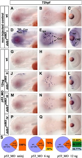
Knock down of p53 rescued the apoptosis phenotype of ddb1m863 mutants. (A-F) Enhanced apoptosis in ddb1m863 (class I phenotype; D-F) compared to wild type siblings (A-C) without p53_MO injection. (G-R) Partial (class II and class III; J-O) and complete (type IV; P-R) rescue of ddb1m863 apoptosis phenotype compared to wild siblings (G-I) after injection of 12 ng p53_MO per embryo. (S) Percentage distribution of mutant and rescued mutant phenotypes. Genotypes were determined by PCR. Class I (orange), mutants showed the severest apoptosis phenotype; class IV (light blue) mutants have the weakest phenotype with only a few of apoptotic cells, similar to their wild type siblings; class II (yellow) and III (green) are intermediate phenotypes. Abbreviations used: tpz, tectal proliferation zone; Cce, cerebellum; ret, retina; Anterior towards the left. Scale bar: 100 µm. |
| Fish: | |
|---|---|
| Knockdown Reagent: | |
| Observed In: | |
| Stage: | Protruding-mouth |