
Fig. S4
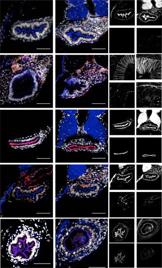
uhrf1pd1092 intestines are morphologically indistinguishable from WT before tnfa elevation at 96 hpf. Shown are confocal images of cross-sections. (A and B) WT and mutant larvae stained for TUNEL. (A′, A′′, B′, and B′′) Typical epithelial morphology in WT (A′) and mutant (B′) larvae. No TUNEL-positive cells in WT (A′′) or mutant (B′′) larvae. WT (C) and mutant (D) larvae stained for pan-cadherin and ZO-1. (C′, C′′, D′, and D′′) Basolateral localization of pan-cadherin in WT (C′) and mutant (D′) larvae. Apical localization (arrows) of ZO-1 in WT (C′′) or mutant (D′′) larvae. WT (E) and mutant (F) larvae stained for mature apical membrane with 4e8 (arrows). (E′, E′′, F′, and F′′) Phalloidin staining in WT (E′) and mutant (F′) larvae. Apical membrane marked by 4e8 staining in WT (E′′) or mutant (F′′) larvae. Enteroendocrine cells in WT (G) and mutant (H) larvae marked by 2f11 (arrows). (G′, G′′, H′, and H′′) Phalloidin staining inWT (G′) and mutant (H′) larvae. 2f11 positive enteroendocrine cells in WT (G′′) or mutant (H′′) larvae. TgBAC(anxa2b-RFP) is a late marker of gut differentiation and is localized apically (arrows) in WT (I) and mutant (J) larvae at 120 hpf. Although the mutant phenotype is present, TgBAC(anxa2b-RFP) is expressed. (I′,I′′, J′, and J′′) Phalloidin staining in WT (I′) and mutant (J′) larvae. Apical TgBAC(anxa2b-RFP) in 120 hpf WT (I′′) or mutant (J′′) larvae. (Scale bars: 50 µm.) |