
Fig. 8
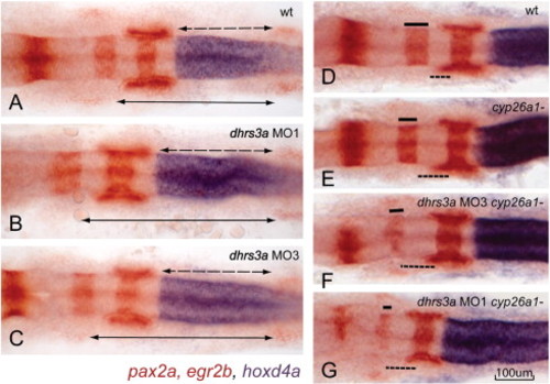
Dhrs3a knockdown effects nervous system patterning. (A–C) in situ hybridization of hoxd4a (blue), pax2a (red in mid-hindbrain boundary, otic vesicles and somites) and egr2b (red in rhombomere 3 and 5) in wt (A), dhrs3a MO1 (B) and MO3 (C)-injected embryos. Dotted arrows indicate the distance from the r6/7 boundary, marked by the anterior limit of hoxd4a expression, to the second somite (indicated by the anterior limit of pax2a expression). Solid arrows indicate the distance from the r3/4 boundary to the second somite. Both distances are longer in dhrs3a knockdown embryos. (D–G) Knockdown of Dhrs3 enhances the posteriorized hindbrain phenotype of cyp26a1 mutant embryos: (D) wt; (E) cyp26a1-/gir mutant; (F, G) cyp26a1-/gir mutant injected with dhrs3a MO3 (F) or MO1 (G). |
| Genes: | |
|---|---|
| Fish: | |
| Knockdown Reagents: | |
| Anatomical Terms: | |
| Stage: | 1-4 somites |
Reprinted from Developmental Biology, 338(1), Feng, L., Hernandez, R.E., Waxman, J.S., Yelon, D., and Moens, C.B., Dhrs3a regulates retinoic acid biosynthesis through a feedback inhibition mechanism, 1-14, Copyright (2010) with permission from Elsevier. Full text @ Dev. Biol.