
Fig. S2
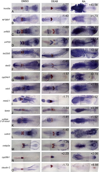
In situ validation of additional microarray hits, continued. In each example, the gene being validated is in blue and egr2b is in red in r3 and r5 as an internal control for the treatments (egr2b expression is eliminated in embryos posteriorized with 0.33 µM RA and r5 is specifically eliminated in embryos anteriorized with 10 µM DEAB). Left column: control embryos treated with 2% DMSO; center column: embryos treated with 10 µM DEAB; right column: embryos treated with 0.33 µM RA. All embryos are shown at approximately 11 hpf with anterior to the left. The numbers in the upper right corners refer to the fold-change in expression level identified on the microarray. |
Reprinted from Developmental Biology, 338(1), Feng, L., Hernandez, R.E., Waxman, J.S., Yelon, D., and Moens, C.B., Dhrs3a regulates retinoic acid biosynthesis through a feedback inhibition mechanism, 1-14, Copyright (2010) with permission from Elsevier. Full text @ Dev. Biol.