
Fig. 5
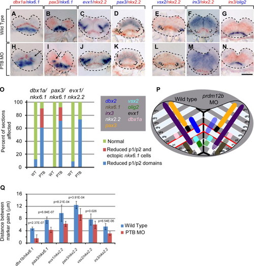
prdm12b Defines the dorsal boundary of nkx6.1 expression and regulates the size of the p1 domain. (A–N) Expression of dbx1a/nkx6.1 (A, H), pax3/nkx6.1 (B, I), evx1/nkx2.2 (C, J), pax3/nkx2.2 (D, K), vsx2/nkx2.2 (E, L), irx3/nkx2.2 (F, M) and irx3/olig2 (G, N) in wild-type (A–G) or PTB MO-injected (H–N) 24 hpf embryos. Panels represent cross sections through the hindbrain with dorsal to the top. (O) Quantification of dorsoventral patterning defects using ~100 sections from 16 embryos for each gene pair. (P) Diagram of dorsoventral gene expression in wild type (left half) and prdm12b MO-injected (right half) embryos. (Q) Quantification of the distance between various gene expression domains using 10 representative sections for each gene pair. Arrows indicate ectopic nkx6.1+ cells and brackets indicate unlabeled tissue between markers. Scale bar is 100 μm. |
| Genes: | |
|---|---|
| Fish: | |
| Knockdown Reagent: | |
| Anatomical Terms: | |
| Stage: | Prim-5 |
| Fish: | |
|---|---|
| Knockdown Reagent: | |
| Observed In: | |
| Stage: | Prim-5 |
Reprinted from Developmental Biology, 390, Zannino, D.A., Downes, G.B., Sagerström, C.G., prdm12b specifies the p1 progenitor domain and reveals a role for V1 interneurons in swim movements, 247-60, Copyright (2014) with permission from Elsevier. Full text @ Dev. Biol.