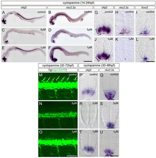
Partial inhibition of Hh signal transduction prior to each step of patterning progression is sufficient to prevent the correct temporal formation of the p3 and p* domains. Panels A-F and M-O show side views of whole embryos; all other panels show spinal cord transverse sections. (A-L) Expression of olig2 (A,C,E; brackets in G,J), nkx2.2a (B,D,F; arrow in H,K) and foxa2 (I,L) at 24 hpf in embryos incubated from 14 hpf in control solution (A,B,G-I) or in cyclopamine at 5 μM (C,D) or 1 μM (E,F,J-L). Note that 1 μM cyclopamine-treated embryos express olig2 (J) but not nkx2.2a (K) and foxa2 (L) in cells abutting the MFP (asterisks). (M-O) Detection of GFP at 72 hpf in Tg(olig2:EGFP) larvae incubated from 30 hpf in control solution (M) or in cyclopamine at 5 μM (N) and 1 μM (O). (P-U) Expression of olig2 (P,R,T) and nkx2.2a (Q,S,U) at 48 hpf in embryos incubated from 30 hpf in control solution (P,Q) or in cyclopamine at 5 μM (R,S) or 1 μM (T,U). Dashed lines indicate dorsal boundary of the nkx2.2a+ domain. Scale bars: 200 μm in A-F; 50 μm in M-O; 20 μm in G-L,P-U.
|
