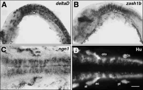
Neurogenesis occurs normally in dtrte370a mutants. (A,B) Lateral views, and (C,D) dorsal views, with rostral to the left, of whole-mounted embryos analyzed either by in situ hybridization (A-C) or by immunohistochemistry (D). The embryos used in each experiment were obtained from a dtrte370a/+ incross, and all embryos exhibited similar labeling indicating that these markers were expressed in a similar fashion in wild-type and mutant embryos. A representative embryo is shown in each case. The asterisk (A,B) indicates the location of the otocyst. (A) At 18 HPF, when branchiomotor neurons are beginning to differentiate, deltaD is expressed extensively in the hindbrain at all dorsoventral levels. (B) At 24 HPF, zash1b is expressed strongly in the hindbrain, especially in dorsoventral columns of cells. (C) At 24 HPF, when branchiomotor neurons are normally still being generated, neurogenin1 is expressed in a broad longitudinal column of cells located medially in the ventral hindbrain. (D) At 24 HPF, the Hu antibody labels cells within longitudinal columns located laterally within the ventral hindbrain. ac, acoustic ganglion; al, anterior lateral line ganglion; oto, otocyst; pl, posterior lateral line ganglion. Scale bar, (A,B) 100 μm, (C,D) 50 μm.
|
