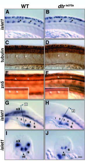
Spinal motor neurons are generated normally in dtrte370a embryos. Panels A-H depict lateral views, with rostral to the left and dorsal up, of the trunk of whole-mounted embryos analyzed either by anti-tubulin (C,D) or zn5 (E,F) immunohistochemistry, or by islet1 in situ hybridization (A,B,G,H). The right-angled arrows in G and H indicate the approximate location and orientation of the transverse sections shown in I and J, respectively, obtained from different embryos. (A) In a 21 HPF wild-type sibling, the two to three isl1- expressing cells (arrowheads) in the ventral spinal cord in every hemisegment are the primary motor neurons. The isl1-expressing cells in the dorsal spinal cord are the Rohon-Beard neurons. (B) In a dtrte370a homozygote, the primary motor neurons (arrowheads) and Rohon-Beard neurons appear normal in number and location. (C) In a 24 HPF wild-type sibling, the primary motor axons exit the spinal cord, with one motor root per hemisegment (arrowheads). (D) In a dtrte370a homozygote, the number and appearance of the primary motor axons exiting the spinal cord (arrowheads) are unaffected. (E) In a 48 HPF wild-type sibling, the zn5-labeled secondary motor neurons are located in the ventral fourth of the spinal cord (arrowheads). Inset depicts a more lateral focal plane showing the secondary motor axons (arrows) exiting the spinal cord and extending ventrally into the somites. (F) In a dtrte370a homozygote, the secondary motor neurons (arrowheads) appear normal in number. However, many secondary motor axons (Inset, arrows) exit the spinal cord at ectopic locations. (G) In a 30 HPF wild-type sibling, the caudalmost nX neurons overlap the rostralmost spinal motor neurons located at the level of somites 2 and 3. (H) In a dtrte370a homozygote, the nX neurons are missing, but the rostralmost spinal motor neurons are still present. (I) Transverse section through the caudal hindbrain in a wild-type sibling shows that the nX neurons and the spinal motor neurons overlap rostrocaudally, but occupy distinct dorsolateral locations. (J) Transverse section through the caudal hindbrain in a dtrte370a homozygote reveals only the rostralmost spinal motor neurons. The apparent difference in isl1 expression in spinal motor neurons between I and J results from the different thicknesses of the sections, which were done by hand. s, somite; nt, notochord. Scale bar, 40 μm.
|
