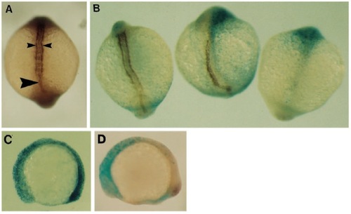
Development of muscle and notochord after inhibition of the FGFR. Dorsal views of embryos at the 14 somite stage, anterior at the top, labelled with antibodies to Ntl and myosin. (A) Normal embryo. The notochord (large arrowhead) is visible in the centre of the embryo running into the tail bud, with bands of myosin staining on either side (small arrowheads). (B) Embryos expressing the dominant negative FGFR showed a range of defects: left embryo has normal myosin-staining and notochord; centre embryo lacks myosin staining on its right side and has a large number of blue cells on this side; right embryo lacks all myosin staining and notochord was only present in the hind-brain. These defects corresponded with a large number of blue cells on both sides of the embryo. (C,D) Distribution of cells inheriting injected RNAs was monitored at 16 hpf using β- gal activity (blue nuclei). (C) Lateral view, anterior to the right, of embryo injected with the control receptor, D50, showing an even distribution of stained cells throughout the anteroposterior axis. (D) Similar embryo injected with the dominant negative receptor, showing stained cells almost exclusively in the head.
|
