
Fig. 6
- ID
- ZDB-FIG-130308-1
- Publication
- Moriarty et al., 2012 - Loss of plakophilin 2 disrupts heart development in zebrafish
- Other Figures
- All Figure Page
- Back to All Figure Page
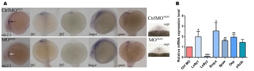
Expression of the cardiogenic marker genes in plakophilin 2 morphant and control morpholino injected embryos. (A) Wholemount in situ hybridization lateral view of nkx2.5 at 24 hpf and bmp4 at 18 somites. Dorsal views of lefty 1 (lft1), lefty 2 (lft2), spaw and oep at 18 somites. nkx2.5 was expressed to the left of the midline in control injected embryos – black arrow; nkx2.5 signal intensity was reduced in morphant embryos, but in the correct location with an expanded domain – white arrow. Expression of lefty 1 was unaffected in morpholino injected embryos while the expression domain of oep was expanded and the intensity of bmp4 and spaw was increased. lefty 2 expression was absent in plakophilin 2 morphant embryos compared to controls. The numbers of embryos with the displayed phenotype were: nkx2.5 control 29/29, nkx2.5 morphant 26/39; lft1 control 44/54, lft1 morphant 47/63; lft2 control 28/34, lft2 morphant 29/35; bmp4 control 34/36, bmp4 morphant 25/32; spaw control 50/57, spaw morphant 39/47; oep control 42/48, oep morphant 36/40. (B) qRTPCR expression of genes involved in cardiac laterality at 18 somites. lefty 1, bmp4, spaw, oep were significantly upregulated and lefty2 expression was significantly decreased in morphant embryos compared to control injected embryos (* P<0.05, ** P<0.005, *** P<0.0001). |
| Genes: | |
|---|---|
| Fish: | |
| Knockdown Reagent: | |
| Anatomical Terms: | |
| Stage Range: | 14-19 somites to Prim-5 |