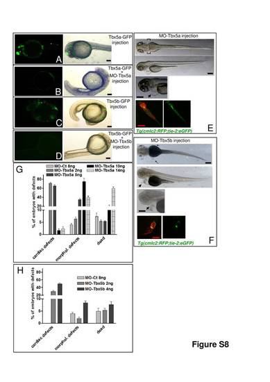
MO-Tbx5a and MO-Tbx5b effectively knockdown the two zebrafish tbx5 isoforms. A-D, 35 pg of pCS2 plasmid expressing GFP fused with MO-Tbx5a or MO-Tbx5b target sequences were injected in one-cell stage embryos in the absence (A and C) or in the presence (B and D) of 1,5 ng of the relative morpholino. Representative fluorescent images of 24 hpf embryos. ~20 embryos for each thesis were analysed. E-F, Tbx5 morphants analysis. Phenotypic analysis of Tbx5a (E) and Tbx5b (F) morphants: 2 ng of MO-Tbx5a, or 4 ng of MO-Tbx5b, were injected in Tg(cmlc2:eGFP) embryos. Phase-contrast images showing pericardial edema (arrowheads) and fin absence (brackets) or presence (arrows); in the bottom right corner of figures E and F, fluorescent images showing heart morphology. Quantification of Tbx5a (G) and Tbx5b (H) morphant phenotypes. The percentage of embryos with the indicate defects was averaged across multiple independent experiments. ~100 embryos for each thesis were analysed. Black scale bars: 100 μm, red scale bars 25 μm.
|
