
Fig. 7
- ID
- ZDB-FIG-120214-5
- Publication
- Malicki et al., 1996 - Mutations affecting development of the zebrafish retina
- Other Figures
- All Figure Page
- Back to All Figure Page
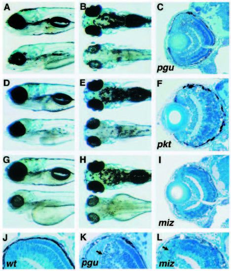
Mutants involving retinal degeneration associated with a general pigmentation defect. Mutant individuals (lower) are shown next to their wild-type siblings (upper). (A,B,C) piegus(pgu)m286. (D,E,F) punktata(pkt)m288. (G,H,I) mizerny(miz)m293. All three mutants have abnormal melanocytes and reduced eye size. Sections through mutant retinae at 3 dpf (C,I) and 5 dpf (F) reveal an excessive amount of cell death. (J,K,L) Higher magnification of the dorsal retina in the wild type (J), pgum286 (K) and mizm293 (L). Cell corpses appear as small, round, intensely staining particles (arrows in K and L). In pktm288 and mizm293 the photoreceptor cell layer has abnormal appearance (F,I). A, D and G show lateral views; B, E and H dorsal views. The dorsal side is oriented up in panels showing lateral views or sections. In panels showing head phenotypes anterior is left. All sections are transverse. |
| Fish: | |
|---|---|
| Observed In: | |
| Stage Range: | Protruding-mouth to Day 5 |