
Fig. s5
- ID
- ZDB-FIG-110622-130
- Publication
- Lupo et al., 2011 - Retinoic acid receptor signaling regulates choroid fissure closure through independent mechanisms in the ventral optic cup and periocular mesenchyme
- Other Figures
- All Figure Page
- Back to All Figure Page
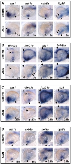
RAR signaling regulates VR/OS and POM genes and is required beyond 18s to maintain their expression. Lateral views of eyes of 31-hpf hybridized embryos treated with DMSO or 10 μM AGN from 3s (A and B) or from 18s or 23 hpf (23h) (C and D), as indicated. (A) AGN treatment causes down-regulation of vax1, net1a, cyb5a, and itgA5 in VR/OS cells (arrowheads). Triangles point to vax1 expression in the ventral forebrain. Arrows point to itgA5 expression in extraocular tissue at the back of the eye. (B) In the control eye, dhrs3a is expressed in VR/OS cells (arrowhead), in the dorsal retina (triangle), and in dorsal POM (arrow). All these domains are down-regulated in the AGN-treated embryo. AGN treatment decreases foxC1a and nlz1 expression in both anterior-ventral (arrowheads) and anterior-dorsal (triangles) POM, although residual foxC1a staining is detectable in a ventro-medial location at the back of the eye (arrows). AGN treatment decreases twist1a expression in dorsal POM only (triangles). (C) Embryos treated with DMSO or AGN from 23 hpf show that AGN treatment causes down-regulation of vax1 and dhrs3a in the VR/OS (arrowheads) and of dhrs3a in the dorsal retina (triangle) and in dorsal POM (arrow). foxC1a and nlz1 expression in both anterior-ventral (arrowheads) and anterior-dorsal (triangles) POM is decreased in the AGN-treated embryos. (D) Embryos treated with DMSO or AGN from 18s or from 23 hpf showing decreased expression of both net1a and cyb5a in the VR/OS (arrowheads) only in the embryos treated with AGN from 18s. |