
Fig. s1
- ID
- ZDB-FIG-110622-126
- Publication
- Lupo et al., 2011 - Retinoic acid receptor signaling regulates choroid fissure closure through independent mechanisms in the ventral optic cup and periocular mesenchyme
- Other Figures
- All Figure Page
- Back to All Figure Page
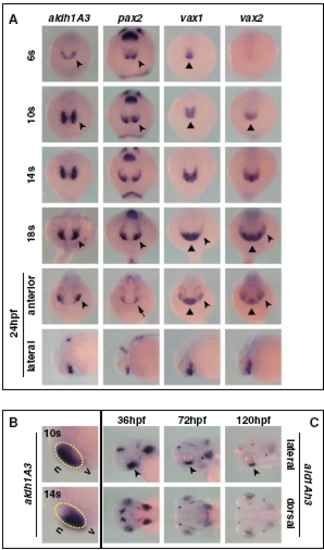
Expression pattern of aldh1A3 during zebrafish development. (A) Frontal/dorsal (6s–18s and 24 hpf, top row) or lateral (24 hpf, bottom row) views of wild-type embryos hybridized with the indicated probes, showing that aldh1A3 is expressed at the right time and place to take part in ventral optic cup morphogenesis by comparison with other known regulators of ventral eye development. During early somitogenesis, expression of aldh1A3 and pax2 becomes detectable in the eye field (arrowheads), whereas vax1 and vax2 expression is limited to the forebrain (triangles). By 14s–18s, all these genes are expressed in the ventral eye (arrowheads), while vax1 and vax2 remain also expressed in the ventral forebrain (triangles). At 24 hpf, aldh1A3, vax1, and vax2 are expressed in the ventral optic cup (arrowheads), while pax2 expression is confined to the optic stalk (OS) (arrow). Similar to previous stages, vax1 and vax2 domains also include the ventral forebrain (triangles). (B) Lateral views of the head region of 10s or 14s embryos, showing that aldh1A3 expression is initially detectable in the nasal (n) part of the eye but becomes localized to the ventral (v) eye as early as 14s. The yellow dashed circles highlight the eye region. (C) Lateral (top row) or dorsal (bottom row) of 36- to 120-hpf embryos, showing that aldh1A3 expression is maintained in the ventral retina (VR)/OS throughout the stages of optic fissure closure (arrowheads). |