
FIGURE
Fig. 2
- ID
- ZDB-FIG-110516-38
- Publication
- Lazic et al., 2011 - Mef2cb regulates late myocardial cell addition from a second heart field-like population of progenitors in zebrafish
- Other Figures
- All Figure Page
- Back to All Figure Page
Fig. 2
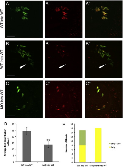
Contribution of wildtype and mef2cb morphant cells to early and late myocardial populations. WT cells transplanted to the WT host margin contributed to both early only (A–A′′) and late (arrowhead) and early (B–B′′) myocardial addition (quantified in E). Morphant cell (0.25 ng) transplanted to the WT host margin contributed only to early myocardial addition (C–C′′). The average number of heart cells found in the WT to WT or MO to WT transplants (D). Green channel (A–C), red channel (A′, B′, and C′), and overlay (A′′, B′′, and C′′). Data shown as mean ± SEM; **P < 0.01. Scale bar represents 50 μm. |
Expression Data
Expression Detail
Antibody Labeling
Phenotype Data
Phenotype Detail
Acknowledgments
This image is the copyrighted work of the attributed author or publisher, and
ZFIN has permission only to display this image to its users.
Additional permissions should be obtained from the applicable author or publisher of the image.
Reprinted from Developmental Biology, 354(1), Lazic, S., and Scott, I.C., Mef2cb regulates late myocardial cell addition from a second heart field-like population of progenitors in zebrafish, 123-133, Copyright (2011) with permission from Elsevier. Full text @ Dev. Biol.