
FIGURE
Fig. S1
- ID
- ZDB-FIG-110516-44
- Publication
- Lazic et al., 2011 - Mef2cb regulates late myocardial cell addition from a second heart field-like population of progenitors in zebrafish
- Other Figures
- All Figure Page
- Back to All Figure Page
Fig. S1
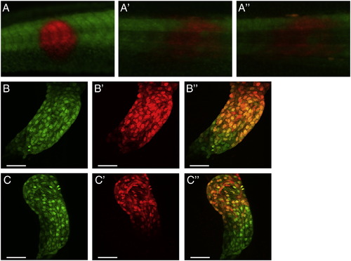
Photoconverted red fluorescing nlsKikGR persists for at least 48 hpf. (A–A′′) Fluorescent microscope images of embryo injected with 100 pg nlsKikGR mRNA at the one-cell stage. A small area of the tail was photoconverted at 24 hpf (A), with red fluorescence readily visible at 48 hpf (A′) and 72 hpf (A′′). (B and C) myl7:nlsKikGR transgenic embryos were photoconverted at 24 hpf and persistence of red fluorescence was ascertained at 48 hpf (B–B′′) and 72 hpf (C–C′′). Green channel (B and C), red channel (B′ and C′), and overlay (B′′ and C′′). Scale bar represents 50 μm. |
Expression Data
Expression Detail
Antibody Labeling
Phenotype Data
Phenotype Detail
Acknowledgments
This image is the copyrighted work of the attributed author or publisher, and
ZFIN has permission only to display this image to its users.
Additional permissions should be obtained from the applicable author or publisher of the image.
Reprinted from Developmental Biology, 354(1), Lazic, S., and Scott, I.C., Mef2cb regulates late myocardial cell addition from a second heart field-like population of progenitors in zebrafish, 123-133, Copyright (2011) with permission from Elsevier. Full text @ Dev. Biol.