
Fig. 2
- ID
- ZDB-IMAGE-110516-38
- Publication
- Lazic et al., 2011 - Mef2cb regulates late myocardial cell addition from a second heart field-like population of progenitors in zebrafish
- All Figures
- Figures for Lazic et al., 2011
|
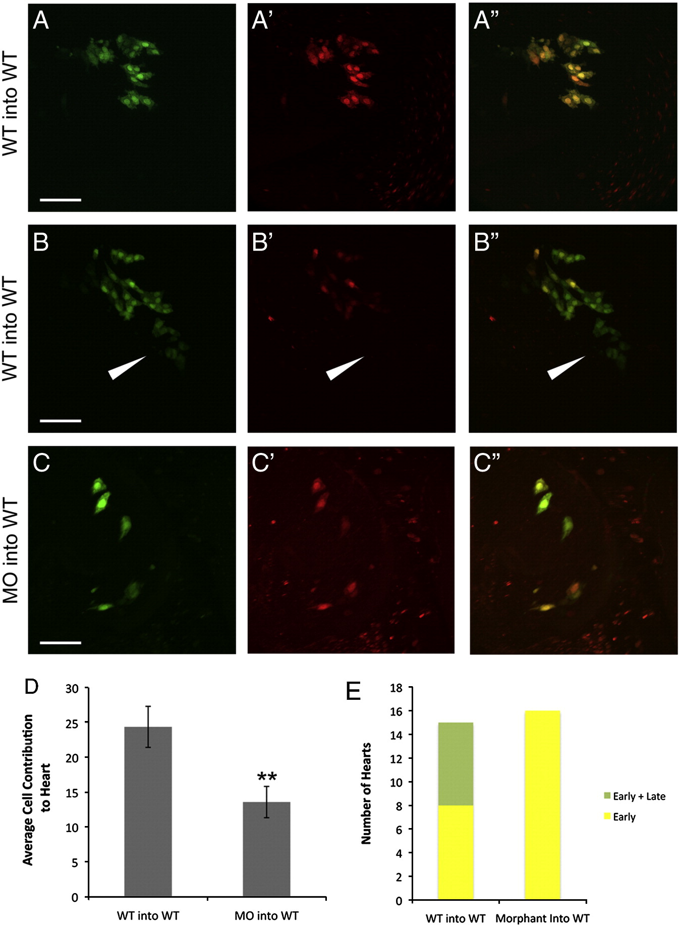
Fig. 2 Contribution of wildtype and mef2cb morphant cells to early and late myocardial populations. WT cells transplanted to the WT host margin contributed to both early only (A–A′′) and late (arrowhead) and early (B–B′′) myocardial addition (quantified in E). Morphant cell (0.25 ng) transplanted to the WT host margin contributed only to early myocardial addition (C–C′′). The average number of heart cells found in the WT to WT or MO to WT transplants (D). Green channel (A–C), red channel (A′, B′, and C′), and overlay (A′′, B′′, and C′′). Data shown as mean ± SEM; **P < 0.01. Scale bar represents 50 μm.
Reprinted from Developmental Biology, 354(1), Lazic, S., and Scott, I.C., Mef2cb regulates late myocardial cell addition from a second heart field-like population of progenitors in zebrafish, 123-133, Copyright (2011) with permission from Elsevier. Full text @ Dev. Biol.