
FIGURE
Fig. S5
- ID
- ZDB-FIG-110516-48
- Publication
- Lazic et al., 2011 - Mef2cb regulates late myocardial cell addition from a second heart field-like population of progenitors in zebrafish
- Other Figures
- All Figure Page
- Back to All Figure Page
Fig. S5
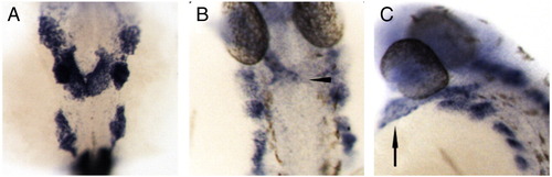
mef2ca marked the heart at 20 and 32 hpf. (A) mef2ca was expressed in the forming heart cone at 20 hpf. Expression is also seen in three bilateral dorsal domains and in the trunk somites. (B and C) At 32 hpf, mef2ca expression was seen throughout the heart tube, with potential late ventricular staining at the arterial pole (arrowhead). Dorsal bilateral and trunk somite staining was maintained. Dorsal view (B) shows ventricular expression of mef2ca, with the lateral view (C) showing expression in the atrium (arrow) as well. |
Expression Data
| Gene: | |
|---|---|
| Fish: | |
| Anatomical Terms: | |
| Stage Range: | 20-25 somites to Prim-15 |
Expression Detail
Antibody Labeling
Phenotype Data
Phenotype Detail
Acknowledgments
This image is the copyrighted work of the attributed author or publisher, and
ZFIN has permission only to display this image to its users.
Additional permissions should be obtained from the applicable author or publisher of the image.
Reprinted from Developmental Biology, 354(1), Lazic, S., and Scott, I.C., Mef2cb regulates late myocardial cell addition from a second heart field-like population of progenitors in zebrafish, 123-133, Copyright (2011) with permission from Elsevier. Full text @ Dev. Biol.