|
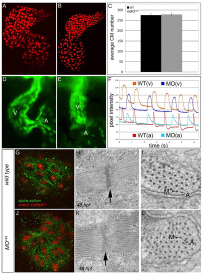
miR-143 is dispensable for proper myocardial and endocardial cell specification, conduction system development and sarcomere assembly. (A,B) Flattened confocal images of 72 hpf WT (A) and MO143 (B) zebrafish hearts carrying a transgene that expresses DsRed2 in cardiomyocyte (CM) nuclei. (C) The average number of total CMs. Error bars represent ± s.e.m. (D,E) WT (D) and MO143 (E) hearts at 48 hpf carrying an flk1::GFP transgene to highlight the endocardium. The ventricle is shown predominantly. (F) Calcium Green fluorescence levels over time in the atria (bottom) and ventricles (top) of 48 hpf WT (blue) and MO143-injected (red) embryos. (G,J) WT and MO143-injected Tg(cmlc2::DsRednuc) embryos (red nuclei) stained with anti-α-actinin (green) to visualize Z-bands at 48 hpf. (H,I,K,L) TEMs of WT and MO143 ventricles at 48 hpf showing Z-disks (H,K, arrows) and sarcomeric bundles (I,L) with organized myosin (M, black arrowheads) and actin (A, open arrowheads) fibers.
|