
Fig. 8
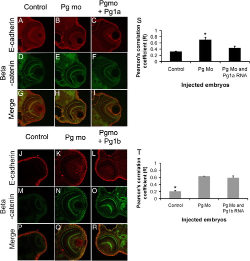
Increased colocalisation of β-catenin and E-cadherin in adherens junctions of plakoglobin morphant embryos. (A–R) Laser-scanning immunofluorescence microscopy images of transverse sections of the eye region of 72 hpf embryos. Dual label fluorescence images with anti-E-cadherin antibody as red and anti-β-catenin antibody as green are shown as (A–F and J–O) single and as (G–I and P–R) merged images. Images are 400x magnification. (G) In control morpholino injected embryos, there was little colocalisation of E-cadherin and β-catenin. (H) By contrast, in plakoglobin morphant embryos there was extensive colocalisation (yellow). (I) This colocalisation was reduced in plakoglobin morpholino and plakoglobin-1a co-injected embryos. (R) However, this colocalisation was still evident in plakoglobin morpholino and plakoglobin-1b co-injected embryos. (S) Quantification of co-localisation of E-cadherin and β-catenin in control, morphant, plakoglobin morpholino and plakoglobin-1a, plakoglobin morpholino and plakoglobin-1b co-injected embryos. There was increased colocalisation of the proteins in morphant embryos compared to control morpholino injected embryos. (S) This co-localisation in co-injected morpholino and plakoglobin-1a RNA injected embryos was similar to control injected embryos. (T) In contrast, levels of co-localisation in plakoglobin morpholino and plakoglobin-1b co-injected embryos were similar to morphant embryos. P < 0.05. Asterisks indicate statistical significance. |
| Fish: | |
|---|---|
| Knockdown Reagent: | |
| Observed In: | |
| Stage: | Protruding-mouth |
Reprinted from Developmental Biology, 327(1), Martin, E.D., Moriarty, M.A., Byrnes, L., and Grealy, M., Plakoglobin has both structural and signalling roles in zebrafish development, 83-96, Copyright (2009) with permission from Elsevier. Full text @ Dev. Biol.