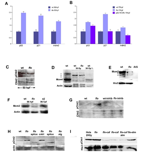
DNA damage response activation in flo mutants. (A,B) Quantitative PCR reveals increased p53, p21, and mdm2 expression in flo mutants. Note that tp53 knockdown in flo (B) abrogates increased p21 and mdm2 expression. (C) Western blot showing comparable levels of phospho-Chk2 in the flo and wild type eye (48 hpf). (D) Western blot showing native and phospho-Mcm4 (*) in the zebrafish wild type (wt), flo intestines before and after γ-irradiation (30 Gy). Note that there is very little native Mcm4 in wt and flo following γ-irradiation (lanes 2 and 3). Phosphatase treatment (λ) of the wt sample from lane 2 dephosphorylates nearly all of the phospho-Mcm4 protein such that only native Mcm4 is present in the sample. (E) Confirmatory Western blot showing reduced chromatin bound Mcm2 in the intestine of 84 hpf flo larvae compared with sibling wt larvae. Far right lane labeled “A/G” shows undetectable levels of Mcm2 and Histone 3 recovered from Ig fraction of the wild type intestinal protein prep prior to anti-histone immunoprecipitation. Presumptive phospho-Mcm2 bands on this gel are denoted by the asterisk (*). (F) Western blot showing reduced Mcm2 in 96 hpf flo larvae, but normal levels in 96 hpf slj larvae compared with control wild type larvae. (G) Western blot showing inhibition of Chk2 phosphorylation in flo larvae treated with the Chk2 inhibitor. (H) Western blot showing specificity of the anti-phospho Chk2 antibody. Phospho-Chk2 levels are elevated in 84 hpf flo larvae but are reduced when injected with splice morpholinos (splice, two independent sets of injections shown) or morpholino designed against the Chk2 translation initiation site (atg) but not larvae injected with vehicle control (cntrl). (I) Western blot showing specificity of the anti-phospho Chk1 antibody: abundant phospho-Chk1 is present in irradiated Hela cells and the non-irradiated 96 hpf flo intestine, but reduced levels are present in the intestine of 96 hpf flo treated with the ATR inhibitor caffeine (10 μM; 15 μM beginning at 84 hpf); the intestine of 96 hpf flo larvae treated with caffeine (10 μM) and a commercially available ATM inhibitor (Sigma Aldrich; 12 μM); but not in the intestine of 96 hpf flo larvae treated with ATM inhibitor alone (12 uM beginning at 84 hpf). His3, anti-Histone 3; A/G, IG fraction recovered following protein A/G precipitation.
|
