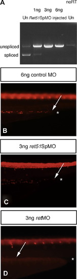
Enteric neurons colonize distal gut regions in the absence of Ret51. (A) RT-PCR analysis reveals that use of a splice blocking morpholino directed against ret51 sequences ret51SpMO on zebrafish embryos, leads to a dose-dependent block of splicing between exon 19 and exon 20, and therefore the loss of the spliced isoform of ret, ret51. RT-PCR on uninjected control fish Un, shows clearly the presence of spliced isoform of ret, ret51. Use of 1ng ret51SpMO leads to a significant reduction of splicing required to generate ret51, whereas use of 3 ng or 6 ng ret51SpMO results in the absence of splicing necessary to generate ret51, and leads to exclusive production of the unspliced form of ret, ret9. The no RT control shows low levels of amplification of contaminating genomic DNA, which as expected, amplifies a band identical in size to the unspliced RNA band. (B–D) Analysis of Hu positive enteric neurons at 4dpf reveals that zebrafish injected with ret51SpMO C show an equivalent distal extent of gut colonization relative to zebrafish injected with a control morpholino (B). In contrast, enteric neurons are absent from distal gut regions in retMO injected zebrafish (D). Asterisk indicates the end of gut tube. Arrows indicate distal most enteric neurons. Lateral views of distal gut are shown. Images for comparison are photographed at the same magnification.
|
