
Fig. S4
- ID
- ZDB-FIG-080325-120
- Publication
- Lachnit et al., 2008 - Alterations of the cytoskeleton in all three embryonic lineages contribute to the epiboly defect of Pou5f1/Oct4 deficient MZspg zebrafish embryos
- Other Figures
- All Figure Page
- Back to All Figure Page
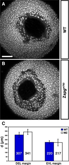
Zygotic Pou5f1 impact on DEL and EVL vegetal closure progression. (A–B) Z-projection of confocal image stacks of a vegetal view of embryos at approximately 90% epiboly stage stained with Sytox green to visualize nuclei. (A) Wild-type (B) Zspgm793. (C) Measurement of the distance of opposing DEL and EVL margins. DEL cells of Zspgm793 displayed an average distance of 341 ± 31 μm and those of wild-type siblings 307 ± 31 μm (p < 0.160) The distance of opposing EVL margins was 220 ± 40 μm in wild-type and 217 ± 19 μm in Zspgm793 (p < 0.951). Embryos resulted from the cross female spg/+x male spg/spg or from the cross female spg/+x male spg/+. The genotypes of embryos were identified after confocal data acquisition by PCR. Scale bar: 100 μm. |
| Fish: | |
|---|---|
| Observed In: | |
| Stage: | 90%-epiboly |
Reprinted from Developmental Biology, 315(1), Lachnit, M., Kur, E., and Driever, W., Alterations of the cytoskeleton in all three embryonic lineages contribute to the epiboly defect of Pou5f1/Oct4 deficient MZspg zebrafish embryos, 1-17, Copyright (2008) with permission from Elsevier. Full text @ Dev. Biol.