
FIGURE
Fig. S9
- ID
- ZDB-FIG-080325-127
- Publication
- Lachnit et al., 2008 - Alterations of the cytoskeleton in all three embryonic lineages contribute to the epiboly defect of Pou5f1/Oct4 deficient MZspg zebrafish embryos
- Other Figures
- All Figure Page
- Back to All Figure Page
Fig. S9
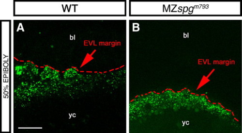
Endocytosis at the YSL-EVL interface. (A, B) Lucifer Yellow soaked 50% epiboly embryos showing vesicle trafficking at the margin. (A) WT; (B) MZspgm793. Z-projection of confocal image stacks; dotted line indicates vegetal margin of EVL. Orientation: animal pole at top. bl: blastoderm, yc: yolk cell. Scale bar: 50 μm. |
Expression Data
Expression Detail
Antibody Labeling
Phenotype Data
| Fish: | |
|---|---|
| Observed In: | |
| Stage: | 50%-epiboly |
Phenotype Detail
Acknowledgments
This image is the copyrighted work of the attributed author or publisher, and
ZFIN has permission only to display this image to its users.
Additional permissions should be obtained from the applicable author or publisher of the image.
Reprinted from Developmental Biology, 315(1), Lachnit, M., Kur, E., and Driever, W., Alterations of the cytoskeleton in all three embryonic lineages contribute to the epiboly defect of Pou5f1/Oct4 deficient MZspg zebrafish embryos, 1-17, Copyright (2008) with permission from Elsevier. Full text @ Dev. Biol.