
Fig. 7
- ID
- ZDB-FIG-080325-110
- Publication
- Lachnit et al., 2008 - Alterations of the cytoskeleton in all three embryonic lineages contribute to the epiboly defect of Pou5f1/Oct4 deficient MZspg zebrafish embryos
- Other Figures
- All Figure Page
- Back to All Figure Page
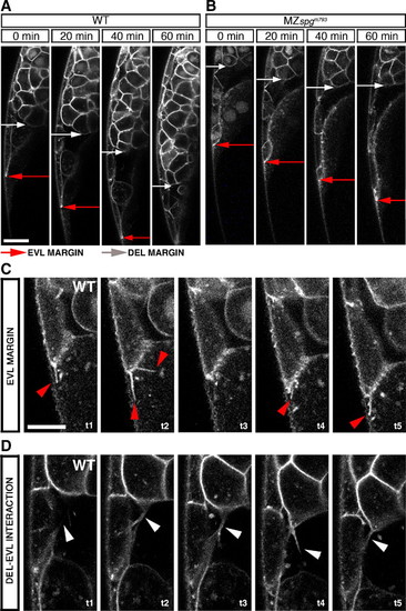
Behavior of cells at the ventral margin. (A–D) Live phenotype observations of the ventral leading edge during a time course of 60 min. Membrane tagged GFP/Sytox green labeled embryos were monitored by confocal z-section time lapse analysis during a time course of 1 h starting at 60% epiboly stage. Red arrows mark the EVL leading edge, white arrows the DEL leading edge. (A) Shows the WT leading edge. (B) Shows the MZspgm793 leading edge. (C) Filopodia formation at the interface between EVL and YSL in a WT embryo (red arrowheads). Δt = 100 s. (D) Filopodia formation between DEL, EVL in WT (white arrowheads). Δt = 250 s. (A, B) Scale bar: 100 μm. (C, D) Scale bar: 50 μm (see also Movies S3, S4, and S5). |
| Fish: | |
|---|---|
| Observed In: | |
| Stage Range: | Shield to 75%-epiboly |
Reprinted from Developmental Biology, 315(1), Lachnit, M., Kur, E., and Driever, W., Alterations of the cytoskeleton in all three embryonic lineages contribute to the epiboly defect of Pou5f1/Oct4 deficient MZspg zebrafish embryos, 1-17, Copyright (2008) with permission from Elsevier. Full text @ Dev. Biol.