
Fig. 3
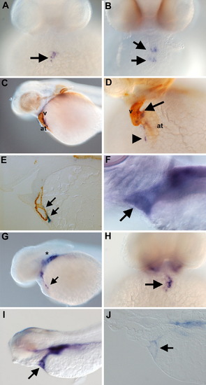
Expression of wt1 and tcf21 in the proepicardial organ. (A) Face view of a 40 hpf embryos stained with wt1. The arrow indicates the wt1-positive proepicardial organ. (B) At 48 hpf wt1 staining is visible as clusters (arrows). (C and D) Lateral and face views of whole-mount embryos co-stained with wt1 (blue) and the anti-myosin heavy chain antibody MF20 (brown). The wt1 clusters localize to the either the atrioventricular junction (arrow in panel D) or the sinus venosus (arrowhead in panel D). (E) Section of 72 hpf embryo stained with wt1 (blue) to label the developing epicardium and MF20 (brown) to label the heart muscle. A streak of wt1-positive cells is visible along the dorsal aspect of the heart (arrows). (F) At 120 hpf, the wt1-positive epicardium surrounds the heart (arrow). (G) Lateral and (H) face views of a 40 hpf embryo stained for tcf21 expression. tcf21 transcripts are located in a cluster of cells between the myoc>-positive epicardium surrounds the heart (arrow). (G) Lateral and (H) face views of a 40 hpf embryo stained for tcf21 expression. tcf21 transcripts are located in a cluster of cells between the myocardium and yolk (arrows) in an identical pattern to wt1. Strong expression of tcf21 is also visible in the developing arches (asterisk in panel G). (I) At 96 hpf, the heart is completely surrounded by tcf21-positive cells (arrow). (J) Sagittal section of a tcf21-stained embryo. As with wt1, the tcf21-positive layer at 96 hpf is confined to the outer epicardial layer (arrow). at: atrium, v: ventricle. |
| Genes: | |
|---|---|
| Fish: | |
| Anatomical Terms: | |
| Stage Range: | Prim-25 to Day 5 |
Reprinted from Developmental Biology, 315(1), Serluca, F.C., Development of the proepicardial organ in the zebrafish, 18-27, Copyright (2008) with permission from Elsevier. Full text @ Dev. Biol.