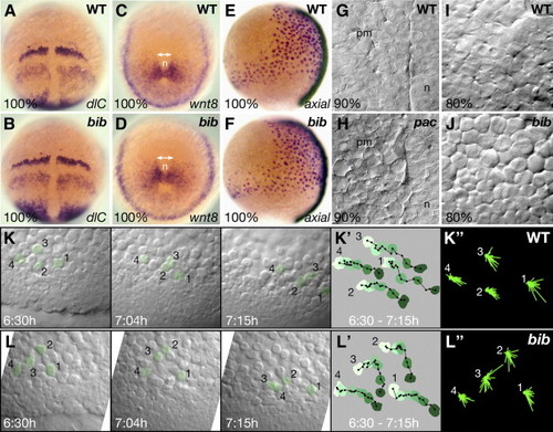
Abnormal cell behavior of the mesoderm in pac and bib mutant gastrulas. (A–D) Whole-mount in situ hybridizations of sibling embryos, double-headed arrows indicate broadened mesodermal territories in the mutant. (E, F) Comparison of axial expression shows normal width of endoderm in mutants. (G–J) High magnification view of the mesodermal layer in live embryos. (G–H) In pac siblings, view of the paraxial mesodermal (pm) territory adjacent to the notochord (n). (I–J) In bib siblings, view of the paraxial mesodermal territory 45° distant from the midline. (K, L) Frames from two simultaneous video timelaspes over a 45-min time period. (K′, L′) Tracings of the net movement for the indicated cells shown in panels K and L; each color is the filled outline of that particular cell for a specific segment in the time-lapse in which time is reflected by change from light green to dark green, filled black circles are the position of the nucleus in that particular cell at 3-min time intervals, black lines are the individual vectors from time point to time point. (K″, L″) Summary roses of the superimposed individual vectors for the indicated cells shown in panels K and L. Note the difference in appearance between mutant cells 2 and 3 and the other cells.
|
