
Fig. 1
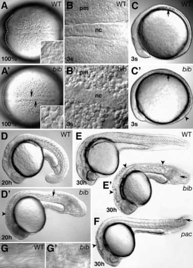
Morphology of the bib phenotype. (A) Dorsal view; arrows indicate kinked mutant notochord, inset shows high magnification of rounded cells in paraxial mesoderm, dotted line outlines ventral portion of anterior neural plate. (B) High magnification of notochord (nc) and paraxial mesoderm (pm). (C) Side view: arrows indicate position of somite furrows; arrowhead indicates flatter mutant tailbud. (D) Side view: arrowhead indicates defects in brain subdivisions; arrow indicates defects in muscle segmentation. Side views of (E) wild-type and bib siblings and (F) pac mutant; arrowheads indicate uneven surface of neural tube, arrows indicate vacuoles in disorganized tail. (G) High magnification of wild type and bib striated muscle. |
| Fish: | |
|---|---|
| Observed In: | |
| Stage Range: | 1-4 somites to Prim-15 |
Reprinted from Developmental Biology, 310(2), Warga, R.M., and Kane, D.A., A role for N-cadherin in mesodermal morphogenesis during gastrulation, 211-225, Copyright (2007) with permission from Elsevier. Full text @ Dev. Biol.