
Fig. 8
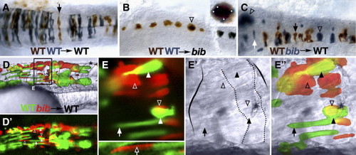
Autonomous and non-autonomous effects of bib in the nervous system and muscle. 24 h mosaic embryos after transplantation of wild-type and/or bib mutant cells into wild-type or bib mutant hosts as indicated. (A–C) Neural transplants into the hindbrain; dorsal view. (A) Arrow indicates typical wild-type neuroepithelial cell. (B) Arrow indicates aggregate of > 10 wild-type cells; inset shows magnification of cluster and white dots indicate individual cells. (C) Arrowheads indicate aggregate mutant cells, white arrow indicates isolated mutant cell, black arrow indicates normal wild-type cell. (D–E) Myotomal transplants into the trunk; lateral view. (D) Low magnification of chimera; dotted lines indicate myotome boundaries; asterisk indicates normal-shaped myotome. (E) Region magnified from panel D, showing UV, white light and composite images; solid arrow indicates typical wild-type fiber, hollow arrow indicates isolated normal-looking mutant fiber, solid arrowheads indicate abnormal wild-type fibers, hollow arrowheads indicate typical mutant aggregates; solid outline indicates abnormal myotome boundaries and dotted outline indicates unusual borders within the myotome. |
Reprinted from Developmental Biology, 310(2), Warga, R.M., and Kane, D.A., A role for N-cadherin in mesodermal morphogenesis during gastrulation, 211-225, Copyright (2007) with permission from Elsevier. Full text @ Dev. Biol.