
Fig. S3
- ID
- ZDB-FIG-070228-9
- Publication
- Murayama et al., 2006 - Tracing Hematopoietic Precursor Migration to Successive Hematopoietic Organs during Zebrafish Development
- Other Figures
- All Figure Page
- Back to All Figure Page
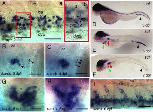
C-myb, scl, ikaros, and runx1 Expression in the Developing Hematopoietic Tissues (A-F,I) lateral views, rostral left; (G,H) ventral views, rostral up; (A) ventral tail at 2 dpf; the area boxed in (a) is displayed enlarged and at a shallower focus in (b), to show that c-myb+ cells are comprised between the caudal artery (ca) and the somite muscle (s) ventral limit (arrows), except for a few in the more ventral CV plexus (cvp) lumen. (B,C) expression in thymus of ikaros at 3 dpf (B) and c-myb at 4 dpf (C); note the single positive cells outside of the thymus anlage, caudalwards (arrowheads); oc, otic capsule. (D-F) From 3 to 7 dpf, scl hematopoietic expression progressively moves from the tail (arrowhead) to the kidney (red arrow); scl+ circulating cells accumulate in the heart (green arrowhead); (G,H) Ikaros (G) and runx1 (H) expression in the 5 dpf kidney, around the pronephric glomerulus (g); (I) Ikaros expression in the 6 dpf tail. Bars, 50 μm |
| Genes: | |
|---|---|
| Fish: | |
| Anatomical Terms: | |
| Stage Range: | Long-pec to Days 7-13 |
Reprinted from Immunity, 25(6), Murayama, E., Kissa, K., Zapata, A., Mordelet, E., Briolat, V., Lin, H.F., Handin, R.I., and Herbomel, P., Tracing Hematopoietic Precursor Migration to Successive Hematopoietic Organs during Zebrafish Development, 963-975, Copyright (2006) with permission from Elsevier. Full text @ Immunity