
Fig. 2
- ID
- ZDB-FIG-070227-5
- Publication
- Murayama et al., 2006 - Tracing Hematopoietic Precursor Migration to Successive Hematopoietic Organs during Zebrafish Development
- Other Figures
- All Figure Page
- Back to All Figure Page
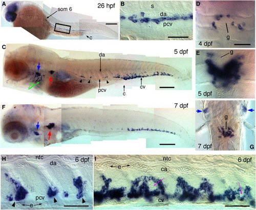
c-myb Expression Reveals the Successive Sites of Larval and Definitive Hematopoiesis Whole-mount in situ hybridization at successive developmental stages. (A) 26 hpf, short after blood circulation onset; position of somite 6 is indicated. (B) Close-up on the area boxed in (A). (C) 5 dpf. (D and E) Ventral views (rostral up) of c-myb+ cells around the pronephric glomerulus (g) at (D) 4 dpf and (E) 5 dpf. (F) 7 dpf. (G) Ventral view (rostral up) of the pronephric region at 7 dpf. (H and I) Higher magnification lateral views (rostral left) of c-myb+ cells in (H) the trunk and (I) the tail, occurring around c-myb- cell clusters (purple arrows). e, primitive erythrocytes; blue arrows, thymus; red arrows, pronephros; green arrows, branchial arches; arrowheads, c-myb+ patches along the PCV. Scale bars represent 200 μm in (A), (C), and (F); 50 μm otherwise. |
| Gene: | |
|---|---|
| Fish: | |
| Anatomical Terms: | |
| Stage Range: | 26+ somites to Days 7-13 |
Reprinted from Immunity, 25(6), Murayama, E., Kissa, K., Zapata, A., Mordelet, E., Briolat, V., Lin, H.F., Handin, R.I., and Herbomel, P., Tracing Hematopoietic Precursor Migration to Successive Hematopoietic Organs during Zebrafish Development, 963-975, Copyright (2006) with permission from Elsevier. Full text @ Immunity