
Fig. 4
- ID
- ZDB-FIG-070228-4
- Publication
- Murayama et al., 2006 - Tracing Hematopoietic Precursor Migration to Successive Hematopoietic Organs during Zebrafish Development
- Other Figures
- All Figure Page
- Back to All Figure Page
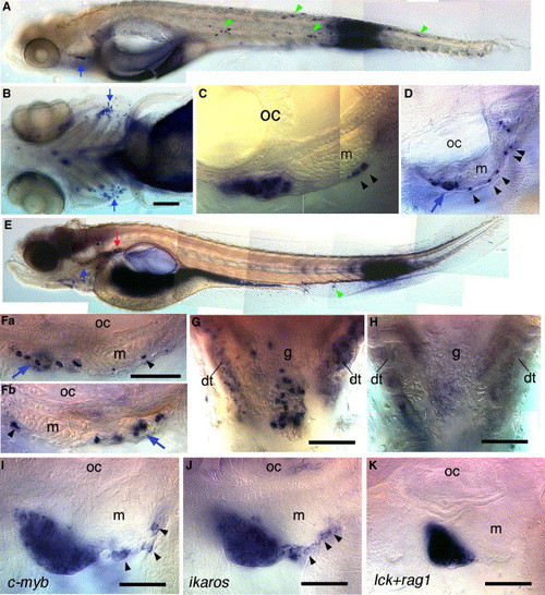
The Tail Contains Future Immigrants to the Thymus and Pronephros (A–C) Caged fluorescein-dextran-injected embryo exposed to UV light in the tail at 48 hpf, fixed and immunostained for fluorescein at 5 dpf. (A) Apart from the large dark spot at the site of uncaging, and the dark gut and yolk (see Experimental Procedures), all spots are labeled cells that emigrated from the initial site of uncaging: to the thymus in large number (arrow), and to various sites as single cells, green arrowheads pointing at those shown in detail in Figure S4 (same in [E]). (B) Ventral view, showing labeling of both thymi (arrows). (C and D) Lateral view of the labeled left thymus (C), and of that of another embryo treated in the same way (D), showing a subepidermal path of labeled cells (arrowheads) to the thymus (arrow). (E–G) Embryo in which fluorescein was laser-uncaged throughout a portion of the caudal hematopoietic tissue at 72 hpf, followed by immunostaining at 5 dpf. The labeled thymi (blue arrows) and pronephros (red arrow) are shown at higher magnification in (F) and (G), respectively; (F) left (Fa) and right (Fb) lateral views; (G) ventral view rostral up. (H) Similar ventral view of a control embryo (caged fluorescein-dextran-injected but not uncaged); in (G), beside the genuinely labeled cells around and above the glomerulus (g), the distal pronephric tubules (dt) show a vesicular labeling (see Experimental Procedures). (I–K) In situ hybridization for (I) c-myb at 5 dpf, (J) ikaros at 6 dpf; (K) lck+rag1 at 5 dpf reveals a path of cells caudal to the thymus for c-myb and ikaros (arrowheads), but not for lck+rag1. m, muscle. Scale bars represent 100 μm in (B) and 50 μm in (F)–(K). |
| Genes: | |
|---|---|
| Fish: | |
| Anatomical Term: | |
| Stage Range: | Day 5 to Day 6 |
Reprinted from Immunity, 25(6), Murayama, E., Kissa, K., Zapata, A., Mordelet, E., Briolat, V., Lin, H.F., Handin, R.I., and Herbomel, P., Tracing Hematopoietic Precursor Migration to Successive Hematopoietic Organs during Zebrafish Development, 963-975, Copyright (2006) with permission from Elsevier. Full text @ Immunity