
Fig. 2
- ID
- ZDB-FIG-060501-3
- Publication
- Miller-Bertoglio et al., 1997 - Differential regulation of chordin expression domains in mutant zebrafish
- Other Figures
- All Figure Page
- Back to All Figure Page
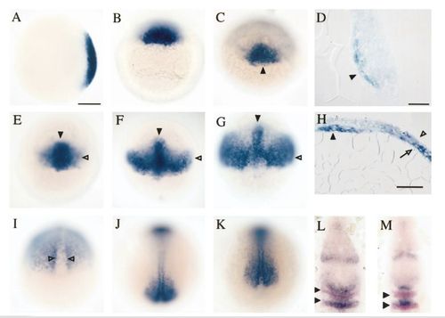
Dynamic expression of chordin in the developing zebrafish. (A, B) Zebrafish chordin was detected at the future dorsal side of the late blastula (sphere stage: A, animal pole view; B, dorsal view). (C, D) By early gastrulation, expression was confined to deep cells in the embryonic shield (arrowhead). D is a sagittal section through the shield at 50% epiboly. (E – H) As gastrulation proceeded, expression was found transiently in the axis (arrowhead) and new bilateral domains of expression appeared (open arrowhead) in all superficial (ectoderm) and deep (mesoderm) cell layers, except the layer closest to the yolk (open arrow, H). E is shield stage at 50% epiboly, F is 30 min after E, and G and H are at 70% epiboly. H is a transverse section through the axis and flanking regions. (I – K) In late gastrulae, chordin was expressed in cells adjacent to the notochord and downregulated in the axis (open arrowheads). Bilateral expression domains persisted in the most posterior region only. I is at 90% epiboly and J and K are at 100% epiboly. (L, M) At late gastrulation, chordin was also expressed in discrete domains in the forebrain, midbrain, and hindbrain (in blue). Double labeling with a probe for krox-20, which is expressed in rhombomeres 3 and 5 (in pink), indicated that chordin expression in the hindbrain is very dynamic, present in different rhombomeres over time. Scale bars = 150 μm (A – C, E – G, and I – M), 25 μm (D), and 50 μm (H). |
Reprinted from Developmental Biology, 192, Miller-Bertoglio, V.E., Fisher, S., Sanchez, A., Mullins, M.C., and Halpern, M., Differential regulation of chordin expression domains in mutant zebrafish, 537-550, Copyright (1997) with permission from Elsevier. Full text @ Dev. Biol.