
Fig. 3
- ID
- ZDB-FIG-060501-4
- Publication
- Miller-Bertoglio et al., 1997 - Differential regulation of chordin expression domains in mutant zebrafish
- Other Figures
- All Figure Page
- Back to All Figure Page
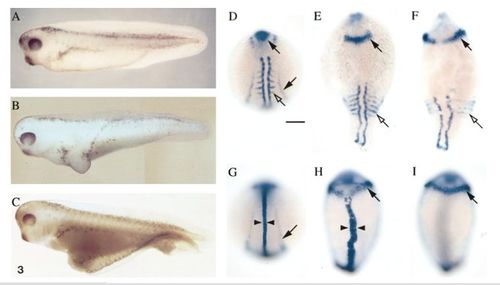
Dorsalizing activity of zebrafish Chordin in Xenopus and zebrafish embryos. (A) Uninjected Xenopus embryo (stage 37/38); (B) Xenopus embryo coinjected with 10 nl of zebrafish chordin and GFP RNA (20 ng/μl) on the ventral side at the two-cell stage; (C) Xenopus embryo coinjected ventrally at the one-cell stage with 5 nl of zebrafish chordin and GFP RNA (20 ng/μl). (D – I) In situ hybridization using pax2 and myoD probes (D – F) or pax2 and ntl probes (G – I) of uninjected zebrafish embryos (D and G) or embryos injected with chordin RNA (E, F, H, and I) at the one- to four-cell stage during segmentation. All injected embryos shown correspond to class d of Table 2. (E, F) Severely dorsalized embryos were characterized by their elongated shape, laterally expanded domain of pax2 expression at the midbrain – hindbrain junction (arrow), and ventrally extending somites (open arrows). (H) In some injected embryos, the ntl expression domain (arrowheads) in the notochord was widened relative to controls (G). D – H are dorsal views. (I) A ventral view of the same embryo as H shows the expansion of pax2 expression in the brain around the entire circumference of the embryo (arrow). Pronephric duct expression of pax2 (arrow, G) was often missing in injected embryos. |
Reprinted from Developmental Biology, 192, Miller-Bertoglio, V.E., Fisher, S., Sanchez, A., Mullins, M.C., and Halpern, M., Differential regulation of chordin expression domains in mutant zebrafish, 537-550, Copyright (1997) with permission from Elsevier. Full text @ Dev. Biol.