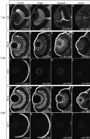
cdh4 knockdown affects differentiation of the retina. A-T: Zebrafish embryos were injected with either standard control morpholino oligonucleotide (MO, A,E,I,M,Q) or cdh4 MO (B-D,F-H,J-L,N-P,R-T). Retinal ganglion cells were visualized at 2 days postfertilization (dpf), using confocal imaging of zn5-immunostained whole-mount embryos. Images are ventral view projections at the level of the optic nerve. Zn5 identifies retinal ganglion cells and their axon processes, among other cells in the developing zebrafish. A: In control embryos, the retinal ganglion cell layer was uniform in thickness and had smooth boundaries. B: The retinal ganglion cell layer was often expanded in cdh4 MO-injected embryos that were only slightly affected. C: In moderately affected embryos, the retinal ganglion cell layer was sparse with irregular boundaries and the optic nerve was thin. D: In severely affected cdh4 knockdowns (D), the retinal ganglion cell layer ranged from nearly absent (D), with only one or two zn5-positive cells and their projections, to altogether absent (not shown). E-T: For Pax6 and zpr-1 immunolocalization, embryos were fixed at 3 dpf (E-L) or at 4 dpf (M-T) and 8 μm horizontal cryosections were cut. Adjacent sections of the same eye (except H, L) were immunostained for Pax6-positive amacrine cells (E-H,M-P) and zpr-1-positive double cone photoreceptors (I-L,Q-T) and visualized by confocal microscopy. cdh4 knockdown images are arranged from left to right as a graded series of slightly affected (F,J,N,R), moderately affected (G,K,O,S), then severely affected (H,L,P,T) embryos. F,H,J,L,N-P,R-T: Amacrine cells and photoreceptor cells fail to differentiate in severe knockdowns (H,L,P,T), but, when present in less affected knockdowns (F,J,N,O,R,S), they were localized in appropriate retinal laminae. Scale bar = 50 μm.
|
