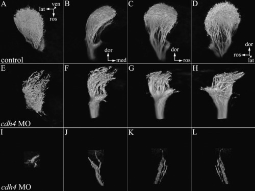
Retinal ganglion cells in 3 days postfertilization (dpf) cdh4 knockdowns arborize only the lateral optic tectum or terminate before reaching the tectum. A-L: Zebrafish embryos were injected with either standard control morpholino oligonucleotide (MO, A-D) or cdh4 MO (E-L). Embryos were fixed at 3 dpf, and retinotectal projections were traced by injecting eyes with the lipophilic dye DiI (1,1'-dioctadecyl-3,3,3',3'-tetramethylindocarbocyanine perchlorate). In both controls and cdh4 knockdowns, retinal ganglion cell axons project toward the contralateral optic tectum. In controls, upon reaching the optic tectum, retinal ganglion cells projections arborized extensively within the tectal neuropil. I-L: In severely affected cdh4 knockdowns, the optic nerve was sparse and failed to reach the optic tectum. E-H: In moderate cdh4 knockdowns, the optic nerve reached the optic tectum, but arborized only the more lateral region. In knockdowns, the nerve fibers were blunt and the fine branching seen in controls was absent. Multiple renderings are shown of each two-photon volume. A,E,I: Dorsal views of the optic tectum, with rostral down and lateral to the left. B,F,J: In the next view, the volumes have been rotated so that we are looking at a rostral view of the fish with dorsal up and lateral to the left. C,D,G,H,K,L: The upright volume renderings of B,F,J have been rotated counterclockwise.
|
