
Fig. 3
- ID
- ZDB-FIG-050531-5
- Publication
- Elworthy et al., 2005 - Phox2b function in the enteric nervous system is conserved in zebrafish and is sox10-dependent
- Other Figures
- All Figure Page
- Back to All Figure Page
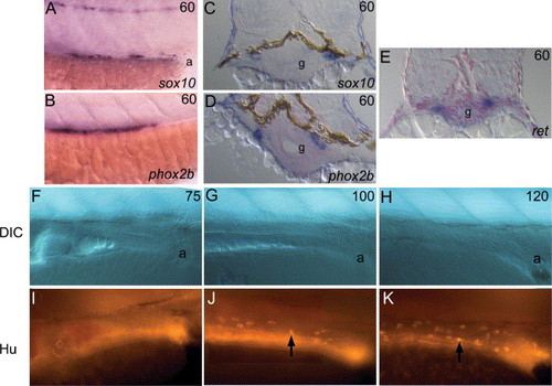
Late embryonic development of the zebrafish enteric nervous system. Lateral views of whole-mount (A, B, F–K) and transverse sections (C–E) of mRNA in situ hybridizations (A–E) or immunofluorescent detection (I–K) and corresponding DIC images (F–H) of markers of ENS progenitors in hindgut region. From 60 hpf, sox10 (A, C), phox2b (B, D) and ret (E) expression is prominent in two chains of ENS progenitors migrating posteriorly along the gut primordium (g). Note that sox10 expression at this stage is seen in cells right up to the anus (a), whereas phox2b (and ret, data not shown) is not expressed this far posteriorly (A, B). In contrast, expression of Hu antigen is not seen in the hindgut at 75 hpf (I), but is seen in increasing numbers of cells at 100 (J) and 120 hpf (K). |
| Genes: | |
|---|---|
| Fish: | |
| Anatomical Term: | |
| Stage: | Pec-fin |
Reprinted from Mechanisms of Development, 122(5), Elworthy, S., Pinto, J.P., Pettifer, A., Cancela, M.L., and Kelsh, R.N., Phox2b function in the enteric nervous system is conserved in zebrafish and is sox10-dependent, 659-669, Copyright (2005) with permission from Elsevier. Full text @ Mech. Dev.