
- Title
-
Zebrafish pigmentation mutations and the processes of neural crest development
- Authors
- Kelsh, R.N., Brand, M., Jiang, Y.J., Heisenberg, C.P., Lin, S., Haffter, P., Odenthal, J., Mullins, M.C., van Eeden, F.J., Furutani-Seiki, M., Granato, M., Hammerschmidt, M., Kane, D.A., Warga, R.M., Beuchle, D., Vogelsang, L., and Nüsslein-Volhard, C.
- Source
- Full text @ Development
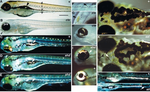
Sixth day phenotypes of decreased cell number (Classes I and II) mutants. Panels show wild-type control sibling together with homozygous individuals for each of cls (A,B,F-H), shd (C-E,I), sal (J,K) and snp (L,M). Chromatophores are prominent in wild-type larvae (four melanophore stripes are labelled, yellow xanthophore pigmentation is clear dorsally, and iridophores are just visible in the eye and the lateral patches (arrowhead)) (A), but absent in cls mutants (B). Note wild-type pigmented retinal epithelium and jaws and arches (arrow) in cls mutants. Occasional chromatophores found in cls include abnormally tiny melanophores (F), but normal xanthophores (G) and iridophores (H). shd mutants (D,E) have fewer iridophores, but where present they look normal. Iridophores in the lateral patches (arrow) and dorsal eye (arrowhead) are highlighted. shd alleles show a phenotypic series: shdty70 (D) is weaker than shdty82(E). An intermediate strength shd allele, shdtm46, shows iridophores over 30% of the outer eye (arrow, I). Ventral views of the yolk sac stripe in snp mutants (M) show gaps in the normally complete (compare L) stripe of iridophores (arrow). The continuous sheet of xanthophores shown in a dorsal view of the head (J) is interrupted in intermediate strength sal alleles (K) by cell-free areas which lack the characteristic granularity and yellow colour of xanthophores (arrow). C-E are dorsolateral views. C-E,I,L and M were photographed with incident light, H with a mix of incident and transmitted light. In this, and all subsequent figures, fish are oriented dorsal up, rostral left, and are photographed with transmitted light, unless otherwise noted. d, dorsal stripe; l, lateral stripe; pre, pigmented retinal epithelium; v, ventral stripe; y, yolk sac stripe. Scale bars, 400 μm (A-E), 75 μm (F), 150 μm (G), 325 μm (H), 250 μm (J,K) and 350 μm (L,M). PHENOTYPE:
|

Sixth day spa mutant phenotype (Class III). Wild-type control (A-C) and spa homozygote sibling (D-F) are shown. Melanophore number is decreased, weakly dorsally (D,E), and more strongly ventrally (arrows in D). Melanophores are abnormally shaped and many are seen as small spots (F, arrowhead) or fragmented (arrow). Xanthophores fill in gaps in the dorsal stripe, giving general yellow colouration in F (compare Fig. 6D). B,C,E and F are dorsal views, of the head and rostral trunk (B,E) or of dorsal stripe melanophores (C,F). Abbreviations as Fig. 2. Scale bars, 400 μm (A,D), 500 μm (B,E) and 125 μm (C,F). PHENOTYPE:
|
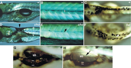
Phenotypes of pigment pattern mutations (Class IV). Wild-type siblings are compared with homozygous mutants for cho (A,B,D,E) on the sixth day, ful on the third day (C,F) and uns on the sixth day (G,H). cho mutants show ectopic melanophores (arrow) in a half collar joining the dorsal and ventral stripes caudal to the ear (D) and have no lateral stripe (arrow, E). ful mutants (F) show laterally displaced dorsal stripe melanophores in the anterior trunk (arrowhead). Close-up view of ventral stripe of wildtype larva (G) shows melanophores covering dorsal surface of swim bladder and large mediolateral extent of lateral patches (flanked by white lines). In uns mutants this region is devoid of melanophores (arrow) and mediolateral extent of lateral patches (white lines) is narrowed (H). C,F are dorsal views of anterior trunk dorsal stripe. Abbreviations as in Fig. 2. e, ear; sb, swimbladder. Scale bars, 300 μm (A,C,D,F) and 150 μm (B,E,G,H). PHENOTYPE:
|
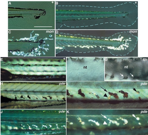
Phenotypes of ectopic chromatophore mutations (Class V). Wild-type siblings are compared with homozygous mutants for mon (A-D), pde (E,H-K) and flh (F,G). mon larvae display on the sixth day large numbers of iridophores extending into the medial fins (C,D), while medial fins (blue outline) are irregular in shape and reduced in area (D). During the pharyngula period, melanophores are normally found in close association with the neural tube in a dorsal position (F), but in flh mutant embryos they completely encircle the neural tube (melanophores on the ventral surface of the tube; arrow, G). On the sixth day, pde larvae show dorsoventrally oriented chromatophores ventral to the notochord and dorsal to the ventral stripe (H, and at higher magnification, in I), which are not present in the wild type (E). Most of these chromatophores look like melanophores under transmitted light, but have large regions (see arrowhead, I) with the appearance of iridophores. When viewed with incident light (J,K), most of these chromatophores (e.g. long arrow, K) look like iridophores. The morphology of these regions strongly implies that the same cell contains both melanosomes and iridosomes. Some of these chromatophores (short arrow, I and K) appear to be pure iridophores. B-D and I-K were photographed with incident light. nt, neural tube. Scale bars, 200 μm (A,C), 400 μm (B,D), 225 μm (E,H,J), 70 μm (F,G) and 110 μm (I,K). |

Sixth day phenotypes of no melanin synthesis and dull iridophore mutations (Classes VI.A and VI.I). Wild-type siblings are compared with homozygous mutants for sdy (A-D) and tnd (E-F). In sdy mutants, gaps (arrow) in the xanthophore pattern correspond to positions of melanophores in wild type (B,D). Both the pigmented retinal epithelium and melanophores are affected. Iridophores are strikingly prominent, as seen in the eye and lateral patches (arrowheads, B). In tnd mutants, iridophores are present in the normal places (arrows, F), but are duller than in wild type. Scale bars, 400 μm (A,B,E,F) and 100 μm (C,D). PHENOTYPE:
|
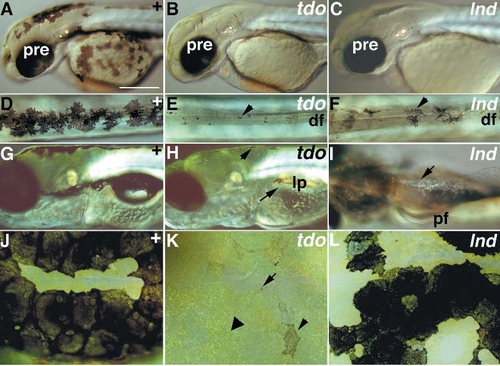
Delayed melanophore differentiation phenotypes (Class VI.B). Wild-type siblings (A,D,G,J) are compared with homozygous mutants for tdo (B,E,H,K) and lnd (C,F,I,L) on the third day (A-F) and the sixth day (G-L). In both tdo and lnd mutants (B and C) the pigmented retinal epithelium is normal, but melanophores are all tiny (tdo, E) or a mixture of tiny and pale cells (lnd, C,F). On the sixth day, tdo mutants now have a few larger, but still pale melanophores (short arrow, H; small arrowhead, K), in addition to tiny melanin spots (arrow, K), although large areas still lack visible melanophores (e.g. none visible around lateral patches: long arrow, H). Xanthophores fill the regions of the dorsal stripe normally occupied by melanophores (large arrowhead, K). In contrast, lnd mutants are now almost fully recovered. Some melanophores are still a little paler (L), and they are still invisible in the yolk sac stripe in some individuals (arrow, I). D-F and J-L are dorsal views of the dorsal stripe in the midtrunk (D-F) or the dorsal head (J-L). I is a ventral view of the trunk. Abbreviations as Fig.2; df, dorsal fin; lp, lateral patch; pf, pectoral fin Scale bars, 250 μm (A-C,G-I) and 100 μm (D-F,J-L). PHENOTYPE:
|
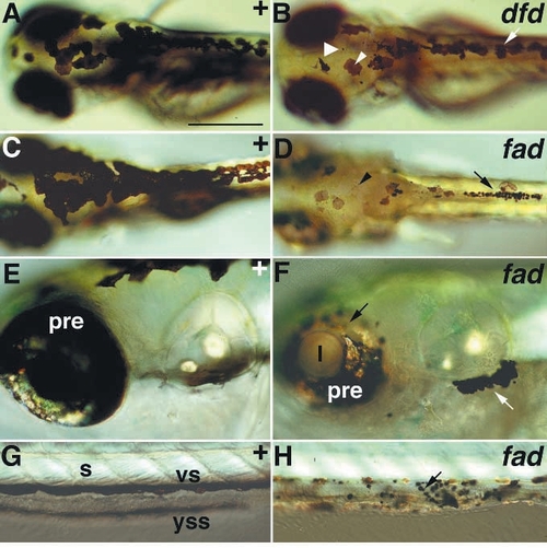
Melanophore degeneration phenotypes (Class VI.C). Wild-type siblings are compared with homozygous mutants for dfd on the fourth day (A,B), and fad on the sixth day (C-H). Abnormal melanophores are first seen mixed in with normal ones (arrow, B) on the fourth day in dfd, as both large, pale cells (small arrowhead, B) and small spots (large arrowhead, B). Abnormal melanophores are first seen on the third day in fad, and by the sixth day the majority are abnormal, being either small and spot-like (arrow, D), pale or apparently fragmented (arrowhead, D). Spot-like melanophores collect abnormally, in piles in the dorsal stripe (arrow, D), in collections ventrolateral to the ear (white arrow, F) and on the hindgut (arrow, H). In fad the pigmented retinal epithelium depigments (D,F) and the eye degenerates (note small size in D,F), although the lens remains normal (F). Abbreviations as Fig. 2; s, somite; l, lens. Scale bars, 350 μm (A-D), 200 μm (E,F) and 125 μm (G,H). |
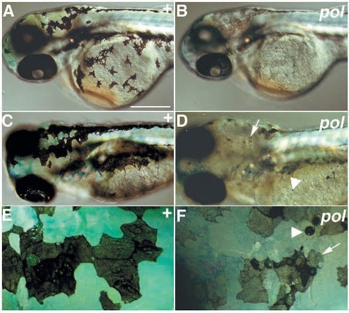
Phenotype of melanophore degeneration and pale xanthophore mutants (Class VI.D). Wild-type siblings are compared with homozygous mutants for pol on day 2 (A,B) and day 5 (C-F). Melanophores on the third day are of normal size, but are very pale (pigmented retinal epithelium is also pale) (B). By the sixth day many melanophores are abnormal in shape (arrow, D,F) and some are small and spot-like (arrowhead, D,F). Xanthophore pigmentation is paler (B,D,F): note that blue colour of xanthophores is due to their taking up methylene blue from the medium. A-D are dorsolateral views; E-F are dorsal views of the head dorsal stripe. Scale bars, 250 μm (A,B), 175 μm (C,D) and 45 μm (E,F). |

Sixth day phenotypes of abnormal melanophore and dull iridophore mutations (Classes VI. E and VI.F). Wild-type siblings (A,B) are compared with homozygous mutants for bry (C,D), frk (E,F) and pun (G,H). In bry mutants, iridophores are slightly dull (arrow, C), and melanophores are pale (D). Unlike bry mutants, mutants in frk and pun combine melanophore degeneration phenotypes (arrows, F and H) with very dull iridophores (E,G). The strength of the iridophore and melanophore phenotypes varies between mutations: pun has a stronger effect than frk, and indeed iridophores can only be seen in the eye (arrow, G). Note in F and H that xanthophores occupy the regions of the dorsal stripe that are without melanophores, thus implying a decrease in melanophore cell number. A,C,E and G are dorsolateral views with incident light; B,D,F and H are dorsal views of the head dorsal stripe. Scale bars, 300 μm (A,C,E,G) and 75 μm (B,D,F,H). PHENOTYPE:
|
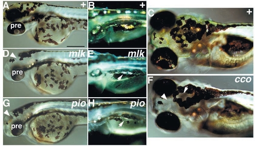
Phenotypes of pale xanthophores and dull iridophores mutations (Class VI. G). Wild-type siblings (A-C) are compared with homozygous mutants for mlk (D,E), pio (G,H) and cco (F) on the third day (A,D,G) and the sixth day (B,C,E,F,H). Xanthophore pigmentation is not visible on the third or sixth days (arrowheads, D,F,G). Iridophores are dull (arrow, E,F) or apparently strongly decreased in number (arrow, H). Eye is small, but normally pigmented, in mlk and pio (D,G). All panels show dorsolateral views. Abbreviations as Fig. 2. Scale bars, 250 μm (A,C,D,F,G) and 325 μm (B,E,H). PHENOTYPE:
|
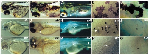
Phenotypes of mutations affecting pigmentation of all chromatophores (Class VI.H). Wild-type siblings (A-E) are compared with homozygous mutants for sbl (F-J) and blc (K-O) on the third day (A,D,F,I,K,N) and the sixth day (B,C,E,G,H,J,L,M,O). Melanophore degeneration begins early (F,I,K,N) and pale (small arrowhead, I), spot-like (arrows, I,N) and fragmenting (large arrowhead, I) melanophores are seen. Melanophore degeneration phenotypes are extreme by the sixth day (G,J,L,O), numbers appear strongly decreased (G,L) and collect abnormally (arrowheads, G). In sbl the eye is small and pale (F), whereas in blc the pigmented retinal epithelium is pale on the third day (K) and largely degenerated by the sixth day (arrow, L). Xanthophores are very pale (F,G,J) or almost unpigmented (K,L,O). Iridophores are dull (arrows, H,M). Panels C,H and M were photographed with incident light. A-C, F-H and K-M are dorsolateral views; D,I and N are dorsolateral views of melanophores on the yolk sac; E,J and O show dorsal views of the head dorsal stripe. Scale bars, 250 μm (A-C,F-H,K-L) and 60 μm (D,E,I,J,N,O). PHENOTYPE:
|

Abnormal chromatophore morphology phenotypes (Class VII). Wild-type siblings (A,G) are compared with heterozygous tin (B), homozygous tin (C), heterozygous pet (E), homozygous pet (F), and homozygous sas (D,H) and uni (I) mutants on the third day (A-F) and the sixth day (G-I). Melanophores are pale and spindly (tin/+, B) or spot-like and uni- or bipolar (tin/tin, C) in tin mutants. Melanophores are pale (pet/+,E) or pale and spindly, but multipolar (pet/pet, F) in pet mutants. sas mutants show small, stellate melanophores with a pale central area (arrowhead, D,H). Melanophores remain small on the sixth day, and xanthophores now have a similar phenotype (arrow, H). uni mutants share the melanophore phenotype (arrowhead, I), but xanthophores are unaffected (I). A-F are dorsolateral views of yolk sac melanophores; G-I are dorsal views of head dorsal stripe. Scale bars, 60 μm (A-F) and 100 μm (G-I). PHENOTYPE:
|

ZFIN is incorporating published figure images and captions as part of an ongoing project. Figures from some publications have not yet been curated, or are not available for display because of copyright restrictions. PHENOTYPE:
|
|
ZFIN is incorporating published figure images and captions as part of an ongoing project. Figures from some publications have not yet been curated, or are not available for display because of copyright restrictions. PHENOTYPE:
|
|
ZFIN is incorporating published figure images and captions as part of an ongoing project. Figures from some publications have not yet been curated, or are not available for display because of copyright restrictions. PHENOTYPE:
|
|
ZFIN is incorporating published figure images and captions as part of an ongoing project. Figures from some publications have not yet been curated, or are not available for display because of copyright restrictions. PHENOTYPE:
|