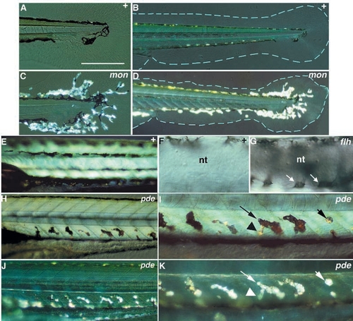
Phenotypes of ectopic chromatophore mutations (Class V). Wild-type siblings are compared with homozygous mutants for mon (A-D), pde (E,H-K) and flh (F,G). mon larvae display on the sixth day large numbers of iridophores extending into the medial fins (C,D), while medial fins (blue outline) are irregular in shape and reduced in area (D). During the pharyngula period, melanophores are normally found in close association with the neural tube in a dorsal position (F), but in flh mutant embryos they completely encircle the neural tube (melanophores on the ventral surface of the tube; arrow, G). On the sixth day, pde larvae show dorsoventrally oriented chromatophores ventral to the notochord and dorsal to the ventral stripe (H, and at higher magnification, in I), which are not present in the wild type (E). Most of these chromatophores look like melanophores under transmitted light, but have large regions (see arrowhead, I) with the appearance of iridophores. When viewed with incident light (J,K), most of these chromatophores (e.g. long arrow, K) look like iridophores. The morphology of these regions strongly implies that the same cell contains both melanosomes and iridosomes. Some of these chromatophores (short arrow, I and K) appear to be pure iridophores. B-D and I-K were photographed with incident light. nt, neural tube. Scale bars, 200 μm (A,C), 400 μm (B,D), 225 μm (E,H,J), 70 μm (F,G) and 110 μm (I,K).
|
