
- Title
-
Sycp2 is essential for synaptonemal complex assembly, early meiotic recombination and homologous pairing in zebrafish spermatocytes
- Authors
- Takemoto, K., Imai, Y., Saito, K., Kawasaki, T., Carlton, P.M., Ishiguro, K.I., Sakai, N.
- Source
- Full text @ PLoS Genet.
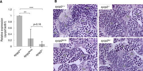
A: Expression of |
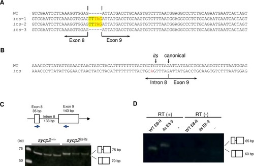
A: cDNA sequences of the exon 8–9 junction of |

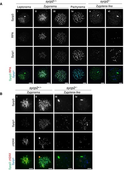
Immunostaining of SC components on wild-type (A), |
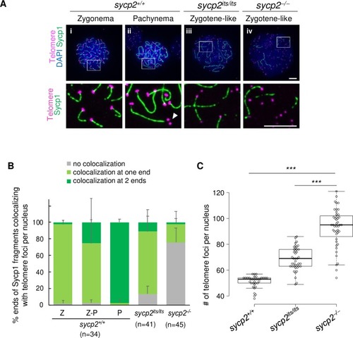
A: Costaining of telomeres and Sycp1 on |
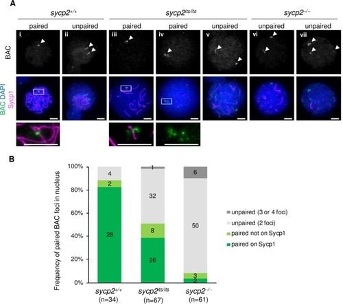
A: Fluorescent in situ hybridization with a BAC probe in |
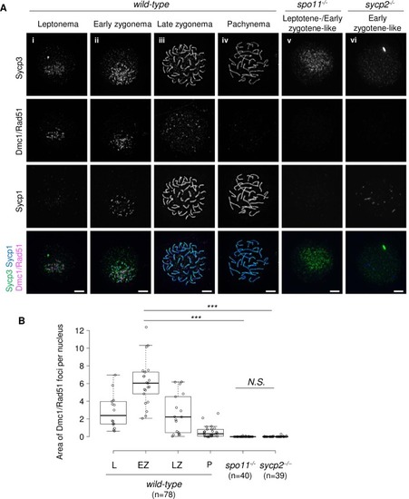
A: Immunostaining of Dmc1/Rad51, Sycp1 and Sycp3 on wild-type (i to iv), |
A: Staining of |

ZFIN is incorporating published figure images and captions as part of an ongoing project. Figures from some publications have not yet been curated, or are not available for display because of copyright restrictions. |
|
ZFIN is incorporating published figure images and captions as part of an ongoing project. Figures from some publications have not yet been curated, or are not available for display because of copyright restrictions. |
|
ZFIN is incorporating published figure images and captions as part of an ongoing project. Figures from some publications have not yet been curated, or are not available for display because of copyright restrictions. PHENOTYPE:
|
|
Unillustrated author statements PHENOTYPE:
|