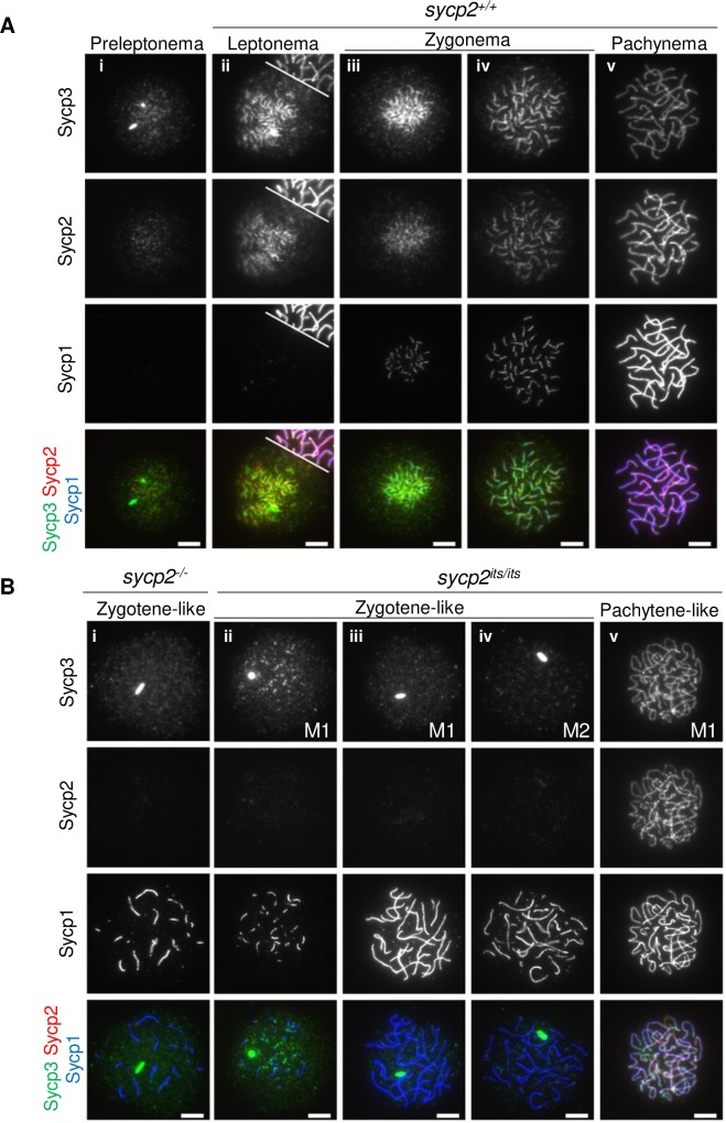
IMAGE

Fig 3

ID
ZDB-IMAGE-200325-60
Genes
Antibodies
Source
Figures for Takemoto et al., 2020
Image
Figure Caption

Fig 3 SC formation is impaired in <italic>sycp2</italic> mutant spermatocytes.

Immunostaining of SC components on wild-type (A), sycp2-/- and sycp2its/its (B) spermatocyte chromosomal spreads. Individual images with anti-Sycp3, anti-Sycp2, or anti-Sycp1 antibodies and a merged image are shown for each nucleus. Wild-type nuclei are staged according to the staining patterns of Sycp3, Sycp2 and Sycp1 (A-i to A-v) [26]. The white line on A-ii indicates a border with another nucleus on the top right. Nuclei from two individual sycp2its/its males (M1 and M2) are shown (B-ii to B-v). Wild-type-like nuclei were observed only among M1 spermatocytes. All spreads were prepared and stained at the same time, and all images were processed in the same manner. Scale bars, 5 μm.

Figure Data
Acknowledgments
This image is the copyrighted work of the attributed author or publisher, and ZFIN has permission only to display this image to its users. Additional permissions should be obtained from the applicable author or publisher of the image. Full text @ PLoS Genet.