IMAGE

Fig 2

ID
ZDB-IMAGE-200325-53
Source
Figures for Takemoto et al., 2020
Image
Figure Caption

Fig 2 Aberrant splicing of <italic>sycp2</italic> mRNA in <italic>sycp2</italic><sup><italic>its/its</italic></sup> testes.

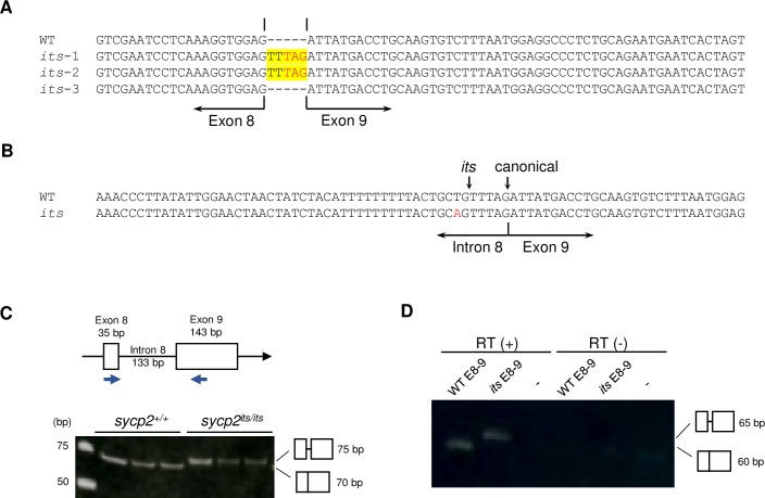
A: cDNA sequences of the exon 8–9 junction of sycp2. The sequences of one sycp2+/+ (WT) and three sycp2its/its (its-1 to its-3) cDNA clones are shown. Two of the three sycp2its/its cDNA clones had a 5-bp insertion (in yellow) containing a premature stop codon (in red letters) at the exon 8–9 junction. B: Genomic sequences of the intron 8-exon 9 junction of sycp2. Sequences from sycp2+/+ (WT) and sycp2its/its (its) testes are shown. The sycp2its allele harbors a T-to-A substitution (in red) upstream of exon 9. The its and wild-type (canonical) splicing sites are indicated with vertical arrows. C: Splicing at sycp2 exon 8-exon 9 was examined by RT-PCR. The target sites of the PCR primers are shown above. Template cDNA was prepared from sycp2+/+ (WT) and sycp2its/its (its) testes from three individual fish. The PCR products were examined on a 20% acrylamide gel. D: Mini-gene splicing assay in the wild-type genetic background. RT-PCR was performed with caudal fin cDNA from transgenic fish with either wild-type (WT E8-9) or its-type (its E8-9) sycp2 mini-genes (S3 Fig). Wild-type fish without mini-genes were used as controls (-). See S3 Fig for more clones and the results of control PCR with a primer pair specific to EGFP.

Acknowledgments
This image is the copyrighted work of the attributed author or publisher, and ZFIN has permission only to display this image to its users. Additional permissions should be obtained from the applicable author or publisher of the image. Full text @ PLoS Genet.