
- Title
-
Expressing acetylcholine receptors after innervation suppresses spontaneous vesicle release and causes muscle fatigue
- Authors
- Mott, M., Luna, V.M., Park, J.Y., Downes, G.B., Epley, K., Ono, F.
- Source
- Full text @ Sci. Rep.
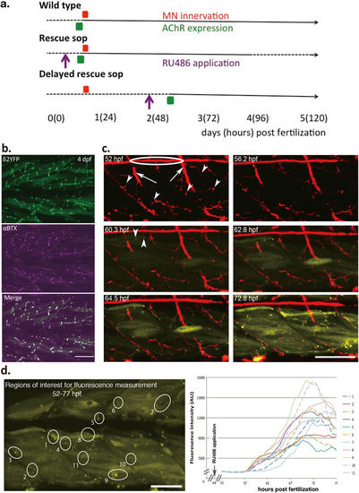
Synapse formation in delayed rescue sop. (a) A summary of the experimental paradigm, showing events related to the synapse development and manipulations along the developmental time course. Numbers indicate days post fertilization (hours post fertilization in parentheses). Dashed lines indicate periods when zebrafish embryos are immotile, and solid lines indicate stages when embryos exhibit mobility. Timing of motor neuron axon innervation (red), AChR expression (green) and RU486 application (purple) are indicated by boxes and arrows. (b) δ2YFP (top), αBTX (middle) and merged (bottom) in the trunk region of a 4 dpf delayed rescue sop are shown. (c) Time-lapse images of axons (red) and AChRs (yellow) in vivo from delayed rescue sop embryos. In the 52 hpf panel, lateral line axons (circled) and motor neuron trunks (arrows) are visualized. Motor neuron terminals are indicated by arrowheads. In the 60.3 hpf panel, the first expressed AChR clusters are detected, indicated by arrowheads. (d) Analysis of fluorescence intensity of identified regions with AChR clusters in delayed rescue sop. Fluorescence from the marked regions of interest (labeled 1–10) was measured and plotted against hours post fertilization. RU486 was applied at 48 hpf, before the measurement of fluorescence started. Scale bars: 50 µm. |

Locomotion of delayed rescue sop. (a) Nerve terminals visualized by anti-SV2 antibody that overlap with the postsynaptic AChRs visualized by αBTX, in wild type and delayed rescued sop embryos at 5 dpf. Scale bar, 50 µm. (b) anti-SV2 antibody staining at 2 dpf, in wild type and in delayed rescue sop before AChR induction. (c) Escape behaviors in wild type and delayed rescue sop at 3 and 5 dpf in response to tactile stimuli. Kinematics for representative traces of 5 larvae are shown for the initial 150 ms of response. Each trace represents a different larva. Body angles are shown in degrees with 0 indicating a straight body, and positive and negative values indicating body bends in opposite directions. Inlaid images of representative larva for each group show superimposed frames of complete escape response and indicate the duration of movement in ms. (d) Maximum turn angle, swim duration and distance traveled were calculated for each group of fish (n = 10 per group) at 3, 4, and 5 dpf. Maximum turn angle is defined as the strongest body angle the larva makes in its initial turn (C-bend) away from the stimulus. Swim duration is defined as the length of time the larva swims following the stimulus until returning to a resting state. Distance traveled is defined as the distance between locations of the head at the start and the end of the swimming. |

Induction of AChR in muscle cells by RU486. a, The scheme of the DNA construct employed for the RU486-inducible system. α-actin promoter drives the expression of a fusion protein4: LexADBD+PRLBD+p65AD. LexADBD is a DNA-binding region of the bacterial LexA protein. PRLBD is a ligand binding domain of the human progesterone receptor. p65AD is the activation domain of the human p65. The δ2YFP gene is downstream of the LexA operator fused to the minimal 35S promoter from Cauliflower Mosaic Virus4. When RU486 binds to the fusion protein, it binds to the LexA operator and drives the expression of δ2YFP. b, The expression of AChRs induced by the RU-486 application. YFP tagged to the AChR was observed in the trunk muscle region. Scale: 1 mm. |