
- Title
-
Zebrafish atoh8 mutants do not recapitulate morpholino phenotypes
- Authors
- Place, E.S., Smith, J.C.
- Source
- Full text @ PLoS One

Atoh8sa1465/sa1465 mutants are morphologically normal, with correct heart looping. a) Structure of the zebrafish atoh8 locus and Atoh8 protein. The positions of the A > T substitution in the atoh8sa1465 allele, and of Lysine 100 in Atoh8 protein, are indicated with asterisks. The red arrowheads mark the positions of each Methionine in the Atoh8 protein, and the basic (orange) and HLH (yellow) domains are indicated. The truncated protein Atoh8K100X is the predicted product of atoh8sa1465. Human ATOH8 contains additional proline-rich (blue) and serine-rich (green) domains. b) Atoh8 Western blot on products from in vitro transcription/translation reactions (TNT Quick) using atoh8WT/WT and atoh8sa1465/sa1465 as templates. Predicted size of Atoh8 = 29.8 kDa. Note that there is a strong nonspecific band at ~31 kDa. The full length membrane can be viewed in S1 Fig. c) qPCR on atoh8WT/WT and atoh8sa1465/sa1465 embryos (hereafter labelled as 'WT' and 'sa1465', respectively). * p = 0.028. d) WT and sa1465 embryos at 5 days postfertilisation, showing normal overall body morphology and swimbladder inflation. e) Confocal z-stacks showing WT and sa1465 embryo heart morphology. EXPRESSION / LABELING:
|
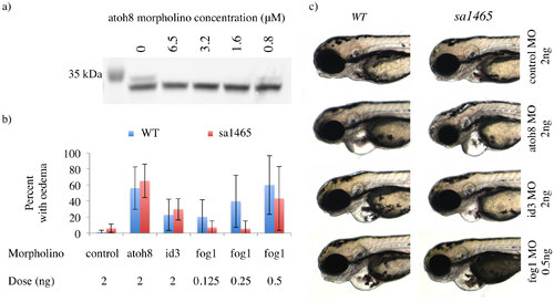
No alteration in response to atoh8 or fog1 knockdown in atoh8sa1465/sa1465 mutants. a) Atoh8 Western blot on products from in vitro transcription/translation reactions (TNT Quick) seeded with atoh8 morpholino at the indicated concentrations. The full length membrane can be viewed in S2 Fig. b) Percentage of WT and sa1465 embryos displaying pericardial oedema at 72 hpf, following injection with the indicated morpholinos. c) Examples of pericardial oedema in WT and sa1465 embryos injected with atoh8, id3, and fog1 morpholinos. Imaged at 72 hpf. PHENOTYPE:
|
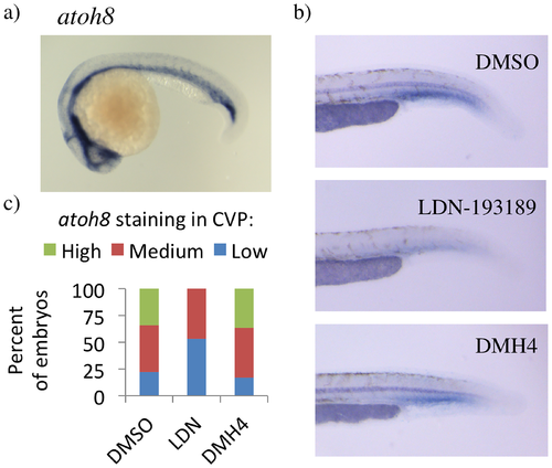
Vascular expression of atoh8, and regulation by Bmps. a) atoh8 expression in 24 hpf Tg(kdrl:eGFP) embryos. b) atoh8 expression in the blood island of 29 hpf embryos. c) Levels of atoh8 staining in caudal vein plexus (CVP) region of 29 hpf zebrafish embryos. Tg(kdrl:eGFP) embryos treated with DMSO, 5 μM LDN-193189, or 10 μM DMH4. Number of embryos: 50 (DMSO), 66 (LDN-193189), 30 (DMH4). |
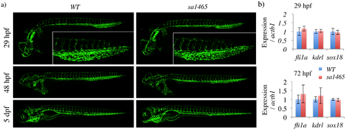
sa1465 embryos have normal vascular patterning and caudal vein plexus formation. a) Confocal z-stacks of WT and sa1465 embryos at the indicated stages. Insets (top) = caudal vein plexus region of 29 hpf embryos. b) qPCR for vascular markers in WT and sa1465 embryos at 29 and 72 hpf. |
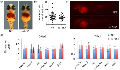
Normal primitive haematopoiesis in sa1465 embryos. a) O-dianisidine staining in 48 hpf WT and sa1465 embryos. b) Number of peroxidase-positive cells present in the distal tail (caudal to the cloaca), in 48 hpf WT and sa1465 embryos. c) Examples of peroxidase staining in 48 hpf WT and sa1465 embryos. d) qPCR for blood markers in WT and sa1465 embryos. PHENOTYPE:
|
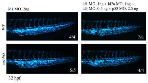
No difference in caudal vein plexus formation between WT and sa1465 embryos injected with id morpholinos. Morpholinos and doses as indicated. Imaged at 32 hpf.
PHENOTYPE:
|