
Image
|
Figure Caption
Fig. 4
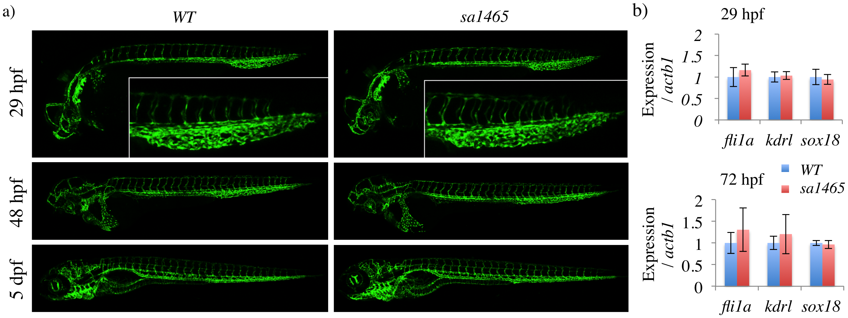
sa1465 embryos have normal vascular patterning and caudal vein plexus formation.
a) Confocal z-stacks of WT and sa1465 embryos at the indicated stages. Insets (top) = caudal vein plexus region of 29 hpf embryos. b) qPCR for vascular markers in WT and sa1465 embryos at 29 and 72 hpf.
Figure Data
Acknowledgments
This image is the copyrighted work of the attributed author or publisher, and
ZFIN has permission only to display this image to its users.
Additional permissions should be obtained from the applicable author or publisher of the image.
Full text @ PLoS One