
- Title
-
Expression pattern of zebrafish rxfp2 homologue genes during embryonic development
- Authors
- Donizetti, A., Fiengo, M., Del Gaudio, R., Iazzetti, G., Pariante, P., Minucci, S., Aniello, F.
- Source
- Full text @ J. Exp. Zool. B Mol. Dev. Evol.

Embryonic gene expression pattern for the three rxfp2 genes by RT-PCR analysis. The different embryonic stages are indicated on top as hours post fertilization. Amplification of rplp0 cDNA was used as a control of RT-PCR sensitivity in the assay. B and O indicate cDNA obtained by retrotranscription of RNA extracted from adult zebrafish brain and ovary respectively. C indicates the negative PCR control reaction lacking cDNA template. |

Spatial localization of rxfp2l transcript during zebrafish late embryogenesis. A: Detail of dorsal region of embryonic brain at 48 hpf. B: Detail of dorsal region of larval brain at 72 hpf. Black arrow indicates the specific hybridization signal. Scale bar: 50 µm. TeO, optic tectum. EXPRESSION / LABELING:
|
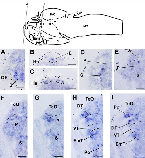
Spatial localization of rxfp2l transcript in the anterior region of zebrafish larval brain. A–I: Transverse sections of hybridized zebrafish larvae (72 hpf) as indicated in the schematic drawing of the zebrafish larval brain. Scale bar: 25 µm. ac, anterior commissure; CeP, cerebellar plate; DT, dorsal thalamus; E, epiphysis; EmT, eminentia thalami; H, hypothalamus; Ha, habenula; MO, medulla oblongata; N, region of the nucleus of medial longitudinal fascicle; oc, optic chiasma; OE, olfactory epithelium; P, pallium; Po, preoptic region; poc, postoptic region; Pr; pretectum; S, subpallium; T, midbrain tegmentum; TVe, telencephalic ventricle; TeO, optic tectum; VT, ventral thalamus. EXPRESSION / LABELING:
|
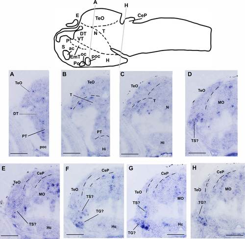
Spatial localization of rxfp2l transcript in the middle region of zebrafish larval brain. A–H: Transverse sections of hybridized zebrafish larvae (72 hpf) as indicated in the schematic drawing of the zebrafish larval brain. Scale bar: 50 µm. ac, anterior commissure; CeP, cerebellar plate; DT, dorsal thalamus; E, epiphysis; EmT, eminentia thalami; H, hypothalamus; Hi, intermediate hypothalamus; Hc, caudal hypothalamus; MO, medulla oblongata; N, region of the nucleus of medial longitudinal fascicle; oc, optic chiasma; P, pallium; Po, preoptic region; poc, postoptic region; PT, posterior tuberculum; S, subpallium; T, midbrain tegmentum; TeO, optic tectum; TG, trigeminal ganglion; TS, torus semicircularis; VT, ventral thalamus. EXPRESSION / LABELING:
|
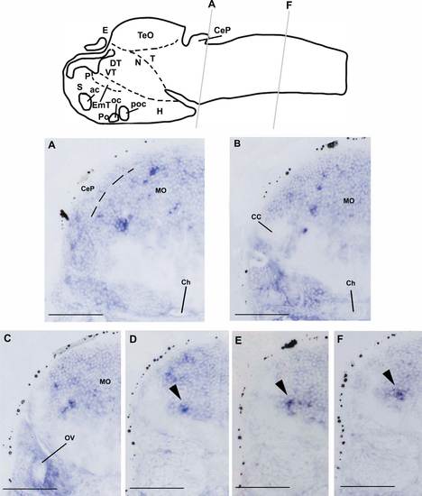
Spatial localization of rxfp2l transcript in the caudal region of zebrafish larval brain. A–F: Transverse sections of hybridized zebrafish larvae (72 hpf) as indicated in the schematic drawing of the zebrafish larval brain. Black arrowhead indicates restricted rxfp2l-expressing cell cluster in the medulla oblongata. Scale bar: 50 µm. ac, anterior commissure; CC, cerebellar crest; CeP, cerebellar plate; Ch, chorda dorsalis; DT, dorsal thalamus; E, epiphysis; EmT, eminentia thalami; H, hypothalamus; MO, medulla oblongata; N, region of the nucleus of medial longitudinal fascicle; oc, optic chiasma; OV, otic vesicle; P, pallium; Po, preoptic region; poc, postoptic region; S, subpallium; T, midbrain tegmentum; TeO, optic tectum; VT, ventral thalamus. EXPRESSION / LABELING:
|

Spatial localization of rxfp2l transcript outside the zebrafish brain. A: Detail of transverse section showing the eye of zebrafish larvae (72 hpf). B: Magnification of inner cell layer as indicated in A. C–F: Detail of transverse sections showing the pharyngeal arches region of zebrafish larvae (72 hpf). Black arrow indicates rxfp2l-expressing cells in the epithelium. Scale bar: 50 µm. a, amacrine cells; b, bipolar cells; Ep, ethmoid plate; gcl, ganglion cell layer; h, horizontal cells; icl, inner cell layer; le, lens; tb, trabeculae. EXPRESSION / LABELING:
|