
Fig. 4
- ID
- ZDB-FIG-151130-15
- Publication
- Donizetti et al., 2015 - Expression pattern of zebrafish rxfp2 homologue genes during embryonic development
- Other Figures
- All Figure Page
- Back to All Figure Page
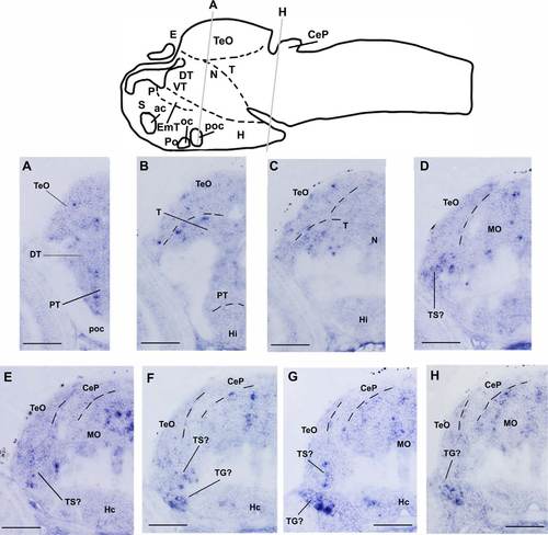
Spatial localization of rxfp2l transcript in the middle region of zebrafish larval brain. A–H: Transverse sections of hybridized zebrafish larvae (72 hpf) as indicated in the schematic drawing of the zebrafish larval brain. Scale bar: 50 µm. ac, anterior commissure; CeP, cerebellar plate; DT, dorsal thalamus; E, epiphysis; EmT, eminentia thalami; H, hypothalamus; Hi, intermediate hypothalamus; Hc, caudal hypothalamus; MO, medulla oblongata; N, region of the nucleus of medial longitudinal fascicle; oc, optic chiasma; P, pallium; Po, preoptic region; poc, postoptic region; PT, posterior tuberculum; S, subpallium; T, midbrain tegmentum; TeO, optic tectum; TG, trigeminal ganglion; TS, torus semicircularis; VT, ventral thalamus. |
| Gene: | |
|---|---|
| Fish: | |
| Anatomical Terms: | |
| Stage: | Protruding-mouth |