
- Title
-
Bmp and Shh Signaling Mediate the Expression of satb2 in the Pharyngeal Arches
- Authors
- Sheehan-Rooney, K., Swartz, M.E., Lovely, C.B., Dixon, M.J., and Eberhart, J.K.
- Source
- Full text @ PLoS One
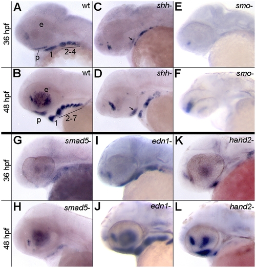
Expression of satb2 requires Hh and Bmp signaling. (A–L) Lateral images of satb2 expression at 36 or 48 hpf. (A–D) Compared to wild-type embryos, in 36 and 48 hpf shha embryos, satb2 expression is reduced in the palatal precursors (arrows in C and D) and throughout the pharyngeal arches. (E,F) Nearly all neural crest cell expression of satb2 appears to be absent in smo embryos. The prominent expression in panel F, is in neural tissue. (G,H) Weak to no expression of satb2 was observed in smad5 mutant embryos at 36 and 48 hpf. (I,J) satb2 expression in edn1 mutants was similar wild-type embryos at both time points analyzed. (K,L) While expression of satb2 was absent throughout the posterior arches of hand2 mutants, the ventral first arch and palatal precursors maintained satb2 expression. p, palatal precursors; e, eye; The arches are numbered in A & C for clarity. |
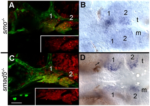
Neural crest cells require the reception of Bmp and Hh signaling to express satb2. (A, C) Lateral views at 36 hpf; (B, D) ventral views at 36 hpf. Both donor and host embryos were fli1:EFGP transgenics to allow for visualization of neural crest cells. (A) Wild-type crest transplanted into the arches of smo mutant embryos are shown in red in the inset. (B) smo+ crest restores the ventral arch expression of satb2 on the side of the transplant (t) as compared to the mutant side of the embryo (m). (C) Lateral views of a 36 hpf smad5 mutant that received a transplant of smad5+ neural crest. The distribution of smad5+ cells (red in inset) is shown. (D) Expression of satb2 is restored in the ventral pharyngeal arch and in the palatal precursors (arrow) on the transplanted side of the embryo. Arches 1 & 2 are numbered in each panel. |

Endodermal signals are required for satb2 expression in ventral arches 2–7. (A) At 38 hpf, satb2 is expressed robustly in the ventral pharyngeal arches and palatal precursors of wild-type embryos. (B) Expression of satb2 in the palatal precursors and ventral arch 1 is retained in sox32 mutants; however, satb2 expression is ablated in the posterior arches. EXPRESSION / LABELING:
|

Expression of shh and satb2 correlate spatially during pharyngeal arch development. (A–D) Dorsal-lateral views of 30 hpf and 32 hpf wild-type embryos. (A) shha expression initiates in the pharyngeal endoderm at 30 hpf while (B) satb2 is not expressed in the craniofacial region at this time. (C, arrows) By 32 hpf shh expression has intensified and can be seen to arc around the presumptive arches. (D, arrows) satb2 expression is initiated in the ventral arch by 32 hpf and lies in close proximity to the presumptive pharyngeal endoderm. EXPRESSION / LABELING:
|
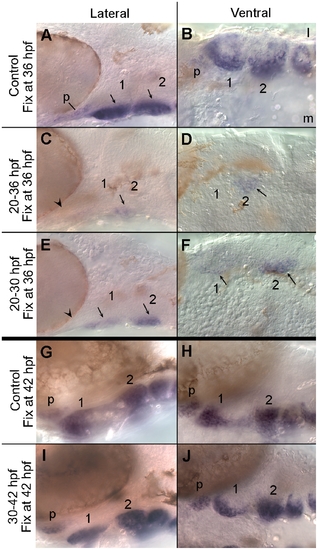
Bmp signaling is required during early arch patterning for the appropriate expression of satb2. (A, C, E) Lateral and (B, D, F) ventral images of 36 hpf embryos labelled with satb2 riboprobe. (A) The ventral region of the pharyngeal arches expresses satb2 robustly. (B) In the ventral arches, satb2 expression is strongest medially. (C, D) Inhibiting Bmp signaling via dorsomorphin from 20 to 36 hpf eliminates the majority of satb2 expression including that in the palatal precursors (arrowhead in C). Only a small population of crest in ventromedial arch 2 expresses satb2 following this treatment (arrows in C & D). (E, F) When Bmp signaling is blocked from 20 to 30 hpf and allowed to recover until 36 hpf, satb2 expression is still greatly reduced. Palatal precursors fail to express satb2 (arrowhead in E) and only ventromedial crest in arches 1 & 2 express satb2 (arrows in E & F). Later inhibition of Bmp signaling does not alter satb2 expression. (G–J) 42 hpf embryos labelled with riboprobe to satb2 in lateral (G, I) and ventral (H, J) views. In both control embryos (G, H) and embryos treated with dorsomorphin from 30 to 42 hpf (I, J), satb2 is strongly expressed by the palatal precursors and in the ventral pharyngeal arches. l, lateral; m, medial; p, palatal precursors. EXPRESSION / LABELING:
|

Continuous Hh signaling is required for satb2 expression. Ventral (A, B, I, J, M, N, Q, R), lateral (C, D, E, F, K, L, O, P, S, T, U, V) and dorsal-lateral (G, H) images of satb2 expression by in situ hybridization are shown in control and cyclopmaine-treated (CYA) embryos. (A, C) In controls at 60 hpf, satb2 is expressed in palatal precursors and in the ventral region of each arch. (B, D) Embryos treated with cyclopamine from 30–60 hpf show a dramatic reduction of satb2 expression in the palatal precursors, pharyngeal arch 1 and 2 as well as complete loss of expression in the posterior arches. (E–H) Compared to controls, embryos treated with cyclopamine from 30 to 36 hpf also show reduction of satb2 expression in the palatal precursors and arches 1 and 2, with a more complete loss of expression in the more posterior arches. (I–L) If, however, embryos are removed from cyclopamine at 36 hpf and allowed to develop for 10 hours, there is a partial recovery of satb2 expression. (M–T) Cyclopamine treatment either from 38 to 42 hpf (M–P) or 44 to 50 hpf (Q–T) results in mild reduction of satb2 expression in the palatal precursors and pharyngeal arches. (U, V) While the maxillary domain is lost in embryos treated with cyclopamine from 6–30 hpf the expression in the ventral arches appears largely intact. CYA, cyclopamine; e, eye; b, brain; p, palatal precursors; pharyngeal arches are numbered in some panels for clarity. |
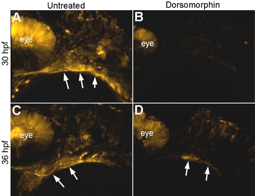
Wash out of Dorsomorphin causes a partial restoration of Bmp signaling. (A) Untreated 30 hpf BRE:mKO2 embryos have rhobust transgene expression in the ventral pharyngeal arches (arrows). (B) Nearly all expression, except for some in the dorsal retina is lost following dorsomorphin treatment from 20–30 hpf. (C) At 36 hpf the expression of the BRE:mKO2 transgene closely resembles that observed at 30 hpf. (D) Embryos treated with dorsomorphin from 20–30 hpf and then washed out of the drug show a partial recovery of BRE:mKO2 expression in the ventral pharyngeal arches (arrows). |