
- Title
-
Identifying the evolutionary building blocks of the cardiac conduction system
- Authors
- Jensen, B., Boukens, B.J., Postma, A.V., Gunst, Q.D., van den Hoff, M.J., Moorman, A.F., Wang, T., and Christoffels, V.M.
- Source
- Full text @ PLoS One
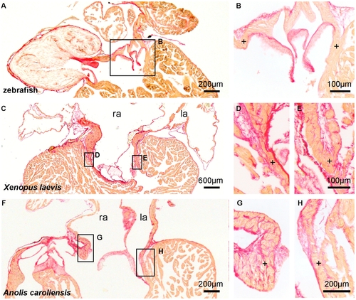
The atrioventricular junction in ectotherms is not interrupted by an insulating plane. Picro-sirius red stain for collagen (red) on 10 μm sections of hearts of adult ectotherms showing the atrioventricular canal to be in full communication (+) with the ventricle. l(r)a, left(right) atrium. |

The phenotype of the slow propagating atrioventricular canal is evolutionary conserved. Numbers in the phylogenetic tree indicate time in millions of years since major splits in tetrapod evolution. (A–E) The hearts of mature ectotherms (blue) and embryonic endotherms (red) maintain complete muscular connection in the atrioventricular canal (avc). (F–J) Markers of fast propagating chamber myocardium (Nppa and Gja5) are absent from the atrioventricular canal (arrowheads). Note that the specimen in H is contracted, obscuring the spongy design otherwise visible. Scale bars in (A–E), 300 μm; (F–J), 100 μm. EXPRESSION / LABELING:
|
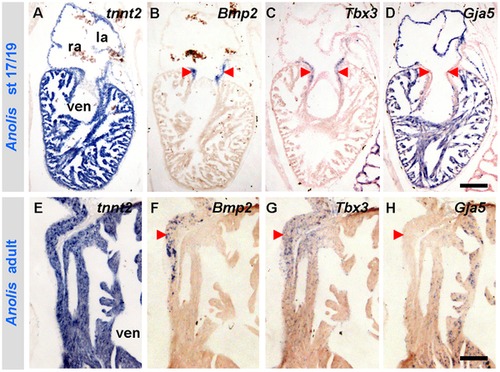
The developmental gene programme of amniotes is maintained in the mature heart of Anolis. (A–D) Stage 17/19 Anolis hearts show complementary expressions of Bmp2 and Tbx3 to Gja5 in the developing myocardial atrioventricular canal (arrowheads). (E–H) The developmental expression of Bmp2, Tbx3 and Gja5 is maintained in the mature myocardial atrioventricular canal (left side shown). la, left atrium; ra, right atrium; ven, ventricle. Scale bars are 100 μm. |
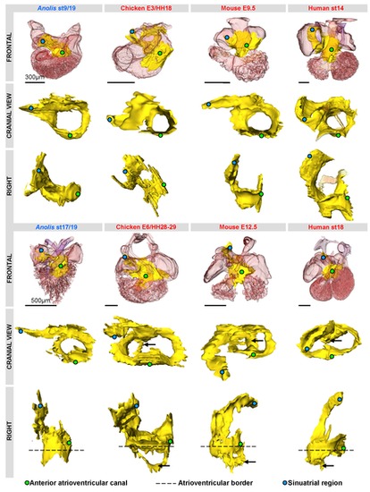
Three-dimensional reconstructions of Tbx3 (yellow) expression in amniotes reveal a shared design. Reconstructions are based on in-situ hybridizations of serial sections, except in human (based on immunohistochemistry, modified from [57], [58]). The Tbx3 domains are strikingly similar in the early phases of chamber formation (upper panel). The Tbx3 expression of the Anolis ventricle is very similar to that associated with ventricular septation (black arrows) in the other amniotes (lower panel). |

Tbx2 expression in the atrioventricular canal of the formed heart of the zebrafish (in-situ hybridization). Nppa is expressed in a complementary pattern. a, atrium; ven, ventricle. EXPRESSION / LABELING:
|

Development of compact walls. The development of the compact walls (Nppa and Gja5 negative) of mammals and birds leaves the trabeculated myocardium (Nppa and Gja5 positive) as a thin inner lining of the ventricular lumina in the fully formed hearts. Nppa is not expressed birds [36]. Scalebars, 100 μm. ivs, interventricular septum; lv, left ventricle. |

Trabeculated ventricles are activated from base to apex. (A, D, G, J) Markers of fast propagating myocardium (Nppa and Gja5) are homogenously expressed in the ventricular trabeculated myocardium from base to apex (ap). (B, E, H) Ventricular activation occurs from base to apex. Early activation is red, late activation is blue. Note that the time-colour coding in panel E is different from that in panels B and H. (K) In species with thick compact myocardium, surface breakthrough of the activation front is earlier in the apical region than in the base. (C, F, I, L) Graphs show the average activation time of the apex and base and the total ventricular activation time. Note that in zebrafish, Xenopus and Anolis, the ventricular base is activated earlier than the apex whereas in mice the ventricular base is activated later than the apex (* Significantly different (one-way ANOVA P<0.05)). n is 3, 6, 9 and 2, respectively. Scale bars in (B, E, H, K) indicate respectively 0.2, 1, 0.5 and 0.1 millimetre. avc, atrioventricular canal; ven, ventricle. EXPRESSION / LABELING:
|
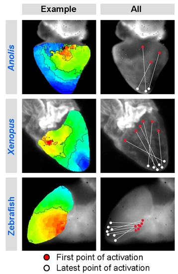
Summary of individual ventricular activation maps. Red marks the earliest epicardial breakthrough of the activation front which was consistently at the ventricular base. Earliest and latest activation (red and white dots respectively) from each specimen is projected onto one specimen. |